
TAME & Musculoskeletal Embolization
TAME for Refractory Patellar Tendinopathy and Hoffa Fat Pad Disease
TAME selectively treats abnormal neovessels that correspond to the painful lesion
This article focuses on TAME/TAE treatment for refractory patellar tendinopathy and Hoffa fat pad disease, not knee osteoarthritis. The more substantial direct evidence for patellar tendinopathy is an 89-patient case series, supplemented by Bow Wang’s two-case report of quadriceps/patellar tendinitis. For Hoffa disease, direct evidence currently consists of a single case from Bow Wang’s team.
The treatment concept is selective microcatheter embolization of arteries supplying abnormal hypervascularity and early venous drainage near the pain generator. Load modification and rehabilitation remain necessary after treatment. Current evidence comes mainly from uncontrolled case series and case reports and does not establish a standard of care.
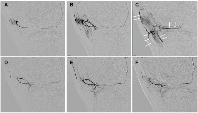
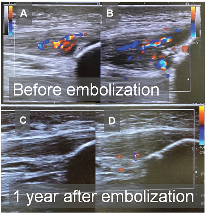
Refractory patellar tendinopathy
Stronger direct evidence: a Level 4 case series with 89 patients completing follow-up, supplemented by one patellar-tendinitis case in Bow Wang’s 2025 two-case JVIR Letter. The signal is encouraging, without a sham or untreated control.
Hoffa fat pad disease
Direct but very early evidence: Bow Wang’s 2022 JVIR case showed pain and functional improvement after two GAE procedures. It remains proof of concept from one patient.
The 89-patient series showed improvement in pain, function, and ultrasound measures, but lacked a control group
2025: 89-patient patellar-tendinopathy series
Among the direct studies currently available, the series by Inui and colleagues has the largest sample. It retrospectively examined recalcitrant patellar tendinopathy treated with TAE from 2017 through 2023. Ninety-eight patients underwent treatment, 9 were lost to follow-up, and 89 were followed for 1 to 7 years, with a mean follow-up of 31.2 months. Mean age was 26.1 years, and the design was a Level 4 case series.
| Outcome | Baseline | 1 month | 3 months | 6 months | Final follow-up |
|---|---|---|---|---|---|
| Pain NRS | 7.6 ± 1.4 | 4.5 ± 2.3 | 3.5 ± 2.4 | 2.7 ± 2.4 | 2.3 ± 2.7 |
| VISA-P | 24.9 ± 15.3 | 43.2 ± 21.6 | 55.2 ± 22.3 | 67.4 ± 24.7 | 74.6 ± 26.2 |
Mean return times were 7.8 weeks for light training and 14.3 weeks for full training; 80 of 89 patients returned to full training within one year. Mean tendon thickness decreased from 9.5 mm to 6.6 mm at final follow-up. No major complication or sonographically visible tendon rupture was observed.
Bow Wang’s team, 2025 JVIR: two cases of quadriceps/patellar tendinitis
In a 2025 JVIR Letter, Bow Wang’s team described two patients with persistent anterior knee pain despite conservative care. MRI showed edema and enhancement in the quadriceps tendon and patellar tendon, respectively; selective angiography demonstrated abnormal neovessels in the corresponding symptomatic regions. Supply arose mainly from the superior patellar and lateral superior genicular arteries in the quadriceps case, and the descending genicular and lateral inferior genicular arteries in the patellar case.
In the patellar case, pain fell from NRS 8/10 to 2/10 within 2 months after one GAE, with resolution of tendon enhancement on follow-up MRI. In the quadriceps case, pain fell from 8/10 to 4/10 after the first treatment but plateaued; repeat GAE was performed 2 months later, followed by a reduction to 2/10 and resolution of edema and enhancement on MRI 2 months after the repeat treatment. No adverse event was reported in either case. The Letter adds diagnostic and technical detail, but a 2/2 outcome cannot estimate population efficacy.
TAME treatment for Hoffa fat pad disease: the 2022 JVIR case
In 2022, Bow Wang’s team reported in JVIR a 32-year-old woman with persistent severe left anterior knee pain, swelling, and inability to bear weight after a traffic accident. Symptoms persisted after surgery and 5 months of corticosteroid treatment and physiotherapy. MRI showed infrapatellar fat-pad hyperintensity and joint effusion. Baseline scores were VAS 9.5/10, WOMAC total 61/96, and WOMAC pain 18/20.
Angiography demonstrated abnormal neovessels in the infrapatellar region supplied by the descending genicular and lateral inferior genicular arteries. VAS fell to 3 one week after the first GAE but then plateaued. Because the vessels had recanalized and the patient sought further relief, repeat GAE was performed at 2 months. One month later, VAS was 1.5, WOMAC total 15, and WOMAC pain 2; VAS was 0 at 2 months, and she remained pain free with an unremarkable range of motion at 6 months. No adverse event was reported.
For a structured imaging differential, see Hoffa’s Fat Pad and Anterior Knee Pain: An MRI Reading Pathway.
How is TAME performed for patellar tendinopathy? From target artery to treatment endpoint
Treatment does not occlude the entire periarticular circulation. A microcatheter is used to identify and selectively treat abnormal vessels that correspond to the painful region. The following steps describe the study protocol; actual strategy depends on lesion location and vascular anatomy.
Ultrasound-guided ipsilateral femoral arterial access and placement of a 3F angiographic catheter.
Assessment of the popliteal, inferior lateral genicular, descending genicular, and inferior medial genicular arteries as appropriate.
Identification of abnormal enhancement and possible early venous drainage near the proximal patellar tendon.
Selective microcatheter delivery of IPM/CS or a short-dissolution gelatin sphere in the study protocol.
Post-treatment angiography to confirm disappearance of abnormal vascular signal and reassess nontarget flow.
Catheter removal, compression, observation, same-day discharge, and planned follow-up of pain, VISA-P, training, and ultrasound.
The IPM/CS concentration, injection increments, and gelatin-sphere preparation were features of this study. Actual material selection depends on regulatory status, vascular anatomy, skin and tendon ischemia risk, and local practice.
When might refractory patellar tendinopathy or Hoffa disease be considered for TAME assessment?
Confirm the true pain generator before TAME
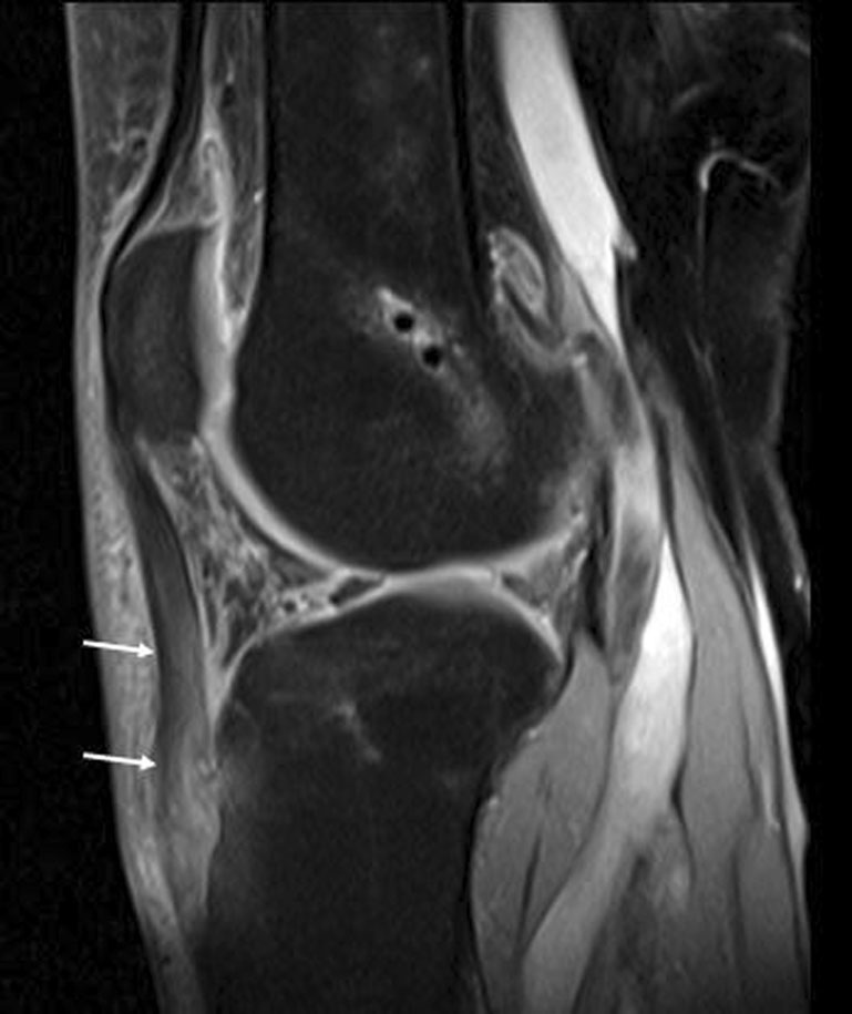
The treatment target must match the clinical pain generator. Anterior knee pain can arise from the patellar tendon, infrapatellar fat pad, patellofemoral joint, plica, bursae, postoperative fibrosis, or another intra-articular lesion. Increased signal in Hoffa’s fat pad on fluid-sensitive MRI is common. It may be the pain generator, a secondary reaction, or an incidental finding.
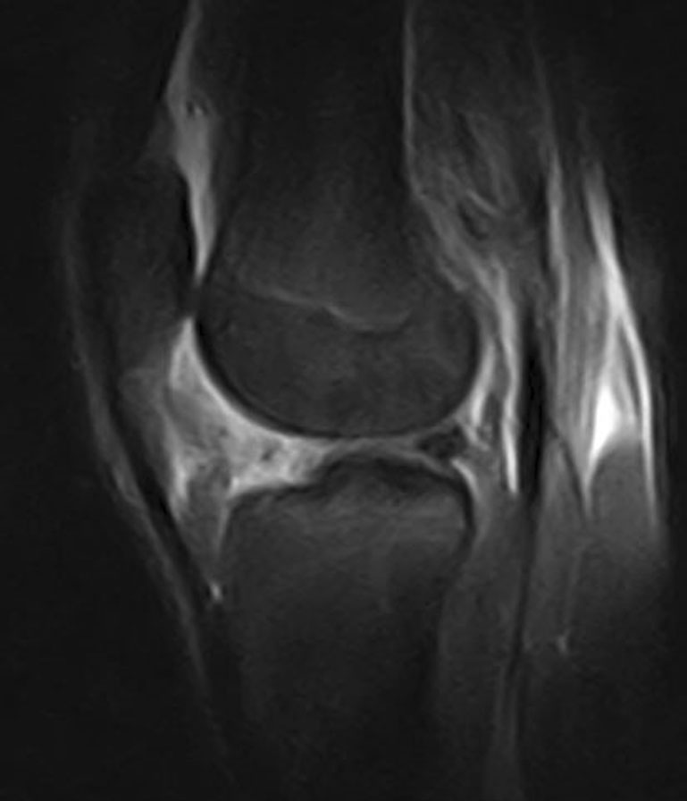
Imaging serves treatment: MRI excludes alternatives, ultrasound follows the tendon, and DSA identifies target vessels
Imaging is not the main subject of this article, but it determines whether TAME has a rational target. MRI evaluates the tendon, Hoffa’s fat pad, patellofemoral joint, and alternative intra-articular causes. Ultrasound can match the point of tenderness while assessing thickness, hypoechogenicity, fiber disorganization, and Doppler flow. Angiography during treatment identifies abnormal local enhancement and early venous drainage.
“No adverse event” in these reports does not mean zero risk
Periarticular knee vessels communicate with cutaneous, neural, muscular, and osseous branches. Potential risks include access-site hematoma, arterial spasm or injury, nontarget embolization with skin discoloration or ischemia, transient sensory symptoms, postembolization pain, and contrast- or medication-related reactions. Because the patellar tendon is a high-load structure, return to sport should follow symptoms, strength, and rehabilitation progress rather than a fixed calendar alone.
Core references and image licensing
- Chen TR, Chang CW, Tai TW, Wang B. Anterior Knee Pain Associated with Hoffa Disease Alleviated with Genicular Artery Embolization: A Case Study. J Vasc Interv Radiol. 2022;33(9):1123–1126.e4. doi:10.1016/j.jvir.2022.05.020. PMID 35688296.
- Sung K, Chen CH, Chen TR, Chen YC, Wang B. Genicular Artery Embolization to Treat Refractory Anterior Knee Pain Associated with Quadriceps and Patellar Tendinitis: Report of 2 Cases. J Vasc Interv Radiol. 2025;36(12):2042–2045. doi:10.1016/j.jvir.2025.08.010. PMID 40816351.
- Inui S, Onishi K, Kochi AT, et al. Transcatheter Arterial Embolization of Abnormal Neovessels for Patellar Tendinopathy: A Safety Evaluation: A Proof-of-Concept Study. Orthop J Sports Med. 2025;13(6):23259671251337093. doi:10.1177/23259671251337093. PMID 40607247.
- Okuno Y, Matsumura N, Oguro S. Transcatheter Arterial Embolization Using Imipenem/Cilastatin Sodium for Tendinopathy and Enthesopathy Refractory to Nonsurgical Management. J Vasc Interv Radiol. 2013;24(6):787–792. doi:10.1016/j.jvir.2013.02.033. PMID 23707086.
- Draghi F, Ferrozzi G, Urciuoli L, Bortolotto C, Bianchi S. Hoffa’s fat pad abnormalities, knee pain and magnetic resonance imaging in daily practice. Insights Imaging. 2016;7(3):373–383. doi:10.1007/s13244-016-0483-8. PMID 27000624.
Image-rights note: This page does not reproduce screenshots of the PDF pages, figures, or captions from either JVIR Letter because a license for public website reproduction has not been confirmed. The displayed images come only from verified licensed sources: Inui et al. Figures 2 and 5 are reproduced unchanged under CC BY-NC-ND 4.0, and Draghi et al. Figures 8 and 14 under CC BY 4.0.
Medical disclaimer: This page provides general medical information and a research summary. It does not replace an in-person consultation or guarantee a specific result. Suitability for TAME requires physician assessment of the history, examination, imaging, previous treatment, and individual risk.
This page is for medical education and academic communication. It does not replace individual diagnosis or treatment advice.
Continue reading this category →