
TAME & Musculoskeletal Embolization
TAME Complications: Prevention and Management in Conventional and Dochu Techniques
Most reported events are mild and reversible, but “uncommon” does not mean negligible
Across the musculoskeletal embolization literature, the events reported most often are transient skin discoloration, access-site hematoma, postembolization pain, and temporary sensory symptoms. Serious events appear uncommon. However, study size, anatomical target, embolic material, and adverse-event definitions vary substantially, so a single percentage cannot represent every TAME procedure.
Safety depends on four linked steps: excluding an incorrect diagnosis and high-risk anatomy before treatment; preventing reflux and nontarget embolization during treatment; actively checking skin, neurologic function, and distal perfusion afterward; and obtaining early imaging when pain or swelling exceeds the expected course.
Separate an expected reaction, an adverse event, and a serious complication
TAME studies commonly use classifications from the Society of Interventional Radiology (SIR) or CIRSE. The defining issue is not the symptom name alone, but whether the event requires additional treatment, prolongs hospitalization, causes permanent harm, or results in death. The table below is a clinical reading aid; formal research reporting should follow the original classification definitions.
| Clinical level | Possible presentation | Practical interpretation |
|---|---|---|
| Expected, transient reaction | Local pain lasting hours to days, mild bruising, or a brief skin-color change | These findings still require documentation and follow-up. Expansion, persistence, or a neurologic or perfusion deficit should not be dismissed as routine. |
| Minor adverse event | Requires outpatient medication, wound care, or additional ultrasound but no admission or invasive intervention | Often reversible, but the duration, treatment, and outcome should be reported rather than summarized only as “no major complication.” |
| Moderate to serious event | Requires an emergency visit, hospitalization, transfusion, surgery, or endovascular repair; causes permanent neurologic injury, tissue loss, or death | Requires prompt recognition and multidisciplinary management and should be addressed during consent even when rare. |
Conventional TAME and ultrasound-guided simplified TAME (Dochu) are not interchangeable
Both approaches attempt to alter abnormal microvascular flow around a painful lesion, but they enter the artery differently and provide different levels of vascular information. Their risk profiles therefore differ.
Conventional TAME
- Uses an arterial sheath and microcatheter, with fluoroscopy and angiography to identify target branches.
- Permits more selective embolization and real-time assessment of abnormal enhancement, anastomoses, and reflux.
- Risks include contrast and radiation exposure, catheter-induced spasm or dissection, nontarget embolization, access-site hematoma, and pseudoaneurysm.
Dochu: ultrasound-guided simplified TAME
- Uses ultrasound-guided percutaneous puncture of an artery near the target and injection of an embolic suspension, without angiography or ionizing radiation.
- Can be faster and less equipment-intensive but is less selective and cannot map collateral or distal distribution as completely as angiography.
- Risks include puncture hematoma, adjacent nerve injury, skin ischemia, allergy, and postembolization pain. Its evidence base is substantially smaller.
Studies of conventional knee, shoulder, or hand embolization cannot serve as direct safety evidence for Dochu. Limited Dochu case experience also does not replace the vascular-anatomy and embolization-safety principles required for catheter treatment.
Each anatomical region has a different risk map
Knee and genicular artery embolization
The adverse event reported most often is transient erythema, livedo, or discoloration caused by embolization of nontarget cutaneous branches. Rates vary widely. In an early 20-patient study, 13 patients (65%) developed skin discoloration and 2 (10%) had paresthesia; all recovered. In another prospective study, 7 participants (18%) developed an approximately 1-cm focal epidermal ulcer. No additional ulcers occurred after the protocol added skin cooling during embolization.45
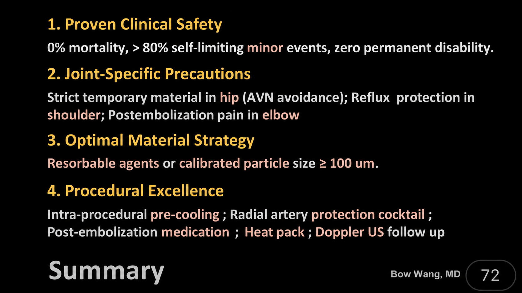
Neurologic symptoms are less common but important. A published case described foot drop after nontarget embolization of a branch near the common peroneal nerve, with recovery by approximately six months. Another case described cutaneous vasculitis after iodized-oil embolization, resolving after three weeks. These reports underscore the need to identify branches toward the fibular neck, skin, and muscle and to control embolic visibility, particle size, injection speed, and reflux.1
Shoulder, hip, foot and ankle, elbow, hand, and wrist
| Region | Common or reported events | Region-specific prevention |
|---|---|---|
| Shoulder | Transient skin erythema, postembolization pain, and access-site hematoma; a rare case of transient cortical blindness has been reported. | Shoulder and cervical vessels have extensive anastomoses. Catheter difficulty, reflux, severe dizziness, or a visual or neurologic symptom should prompt immediate cessation and assessment. A meta-analysis of 329 shoulders recorded 94 events, 92.6% of which were mild.210 |
| Hip | Skin discoloration, postembolization pain, hematoma, and posterior-thigh numbness; femoral-head osteonecrosis is a major theoretical harm to prevent. | Terminal retinacular vessels supply the femoral head. The lecture recommends temporary, rapidly resorbable agents. This is an important expert precaution, but material selection remains individualized and should evolve with evidence. |
| Foot and ankle | Access hematoma, spasm, dissection, distal arterial occlusion, skin ischemia, or ulceration | Vessels are superficial and the tissue compartments are tight. Reports of pedal or tibial access describe local hematoma in approximately 2.5%–17%, usually without additional intervention.7 |
| Elbow | Postembolization pain may last longer. | In a 2025 multiregion study, pain beyond seven days occurred in 5.8%–22.1% depending on the agent, with the elbow showing the greatest frequency and duration. This was observational, not a randomized material comparison.9 |
| Hand and wrist | Transient fingertip pallor, intrinsic-muscle weakness, or dull pain; brachial dissection and pseudoaneurysm have also been reported. | Digital circulation and nerves are closely packed. In a 58-patient hand study, 5 patients (8.6%) developed temporary weakness or dull pain that resolved within 7–10 days.11 |
Embolic material is not a simple choice between “more durable” and “more precise”
Imipenem/cilastatin crystal suspension (IPM/CS), rapidly or temporarily resorbable gelatin sponge, resorbable microspheres, and permanent microspheres have different physical behavior. A 2026 knee review of 22 studies, 633 patients, and 719 knees found no significant pain or functional difference between IPM/CS and permanent particles. Two sham-controlled trials also found no significant efficacy advantage over sham treatment. One permanent-particle case series reported asymptomatic osteonecrosis. These findings support balancing evidence, target-vessel anatomy, and ischemic risk rather than treating any one material as an established universal standard.3
In the Finas shoulder study, complications occurred in 11 of 15 patients (73.3%) in the microsphere group and 3 of 14 (21.4%) in the IPM/CS group. The sample was small and nonrandomized, and multiple events could occur in one patient; the result does not establish that all microspheres are intrinsically more dangerous.8
The access route is part of the procedure’s risk
Radial, ulnar, brachial, femoral, and pedal access each has advantages and limitations. Selection should account for vessel diameter, the sheath-to-artery ratio, ultrasound visibility, compression conditions, and alternative access—not proximity to the target alone.
One patient developed severe forearm and finger pain with numbness in a median-nerve distribution the day after treatment. Ultrasound and CT angiography confirmed a pseudoaneurysm, and symptoms gradually improved after vascular surgical repair. Delayed or progressive pain, swelling, and neurologic symptoms should not be managed with analgesics alone.
Allen or Barbeau testing should not be treated as a single threshold that predicts all hand ischemia. The 2018 AHA statement and 2020 SCAI document no longer recommend excluding radial access solely because of an abnormal test in the coronary-intervention setting. That evidence comes from cardiovascular practice and should be integrated cautiously with vascular ultrasound, device size, and the actual TAME anatomy rather than copied without context.1314
Dochu’s convenience comes with less vascular information
Dochu can be performed rapidly under ultrasound without ionizing radiation and may reduce equipment and time requirements. It is also a less selective injection without angiographic confirmation of distal cutaneous branches, hazardous anastomoses, or reflux. Current publications consist mainly of technical descriptions and limited case experience. Dochu should not be described as standard treatment, and its risk should not be assumed to be lower than conventional TAME.
| Dochu-related issue | Prevention focus | Management direction |
|---|---|---|
| Hematoma or extravasation | Keep the needle tip visible in the lumen, confirm Doppler flow before and after injection, and compress according to needle size and location. | Local compression and follow-up; obtain ultrasound when swelling, pain, or pulsatility persists to exclude pseudoaneurysm or occlusion. |
| Persistent pallor or discoloration | Mix the suspension thoroughly, avoid repeated puncture and excessive injection, and monitor distal perfusion. | Provide warmth and assess perfusion immediately. Vasodilator or antiplatelet medication requires physician-directed, individualized use; patients should not self-medicate. |
| Skin ulceration | Avoid overembolization and temporarily avoid high-impact activity. Use greater caution in Raynaud phenomenon or when distal perfusion is uncertain. | Begin wound care early and refer to dermatology, plastic surgery, or vascular specialists when appropriate. |
| Adjacent nerve puncture | Map nerve, artery, and needle path before puncture. The wrist, anterior neck, and medial thigh require attention beyond the vessel alone. | Persistent numbness, weakness, or neuropathic pain requires prompt neurovascular examination and ultrasound rather than repeated blind injection. |
| Allergy or drug eruption | Review contrast, antibiotic, and medication allergies and prepare for acute hypersensitivity treatment. | Treat according to severity. Dyspnea, facial or tongue swelling, or a systemic reaction requires emergency care. |
The source lecture uses “500 mg IPM/CS in 10 mL contrast, no more than 2 mL per vessel” as a practical reminder against overembolization. For that formulation, this corresponds to approximately 100 mg per vessel. This concentration and limit have not been validated in a prospective trial and must not be transferred to another concentration, material, anatomical region, or patient.
The diagnosis should also be reconsidered before treatment. Acute trauma or infection, tumor such as a glomus tumor, Raynaud phenomenon, chronic cold-type complex regional pain syndrome, or pain dominated by widespread allodynia and central sensitization may require a different diagnostic or therapeutic priority. These examples are clinical concerns for professional assessment, not an absolute self-screening contraindication list.
A safety checklist before, during, and after treatment
Before treatment
Confirm the diagnosis and pain generator. Exclude infection, acute trauma, tumor, and major distal-perfusion problems. Review renal function, allergy, coagulation, and medication. Map access and hazardous anastomoses with ultrasound or MRA when appropriate.
During treatment
Maintain selectivity, inject slowly, and watch for reflux. Use region-appropriate skin protection. Stop for resistance, pain, dizziness, visual, or neurologic symptoms. In Dochu, keep the needle tip and target vessel continuously visible.
After treatment
Compare bilateral pulse, temperature, color, sensation, and strength. Mark hematoma margins and document skin change. Provide explicit warning signs and contact instructions. Arrange Doppler follow-up when indicated rather than monitoring pain score alone.
What can be monitored, and what requires escalation?
| Symptom | First assessment | Escalation signs |
|---|---|---|
| Local or increasing pain | Assess timing, location, and consistency with the expected course; examine color, temperature, pulse, sensation, and strength. | Rapid progression, uncontrolled pain, firm swelling, pallor, coldness, numbness, or weakness. |
| Erythema or discoloration | Mark the area, photograph progression, and check blanching and capillary refill. | Blister, ulcer, dark purple or black change, expansion, or persistent hypoperfusion. |
| Access-site mass or bruising | Apply compression, assess pulsatility and bruit, and perform color Doppler. | Continued expansion, falling hemoglobin, severe pain, nerve compression, or suspected pseudoaneurysm. |
| Numbness, weakness, or foot drop | Perform a complete neurologic and vascular examination and distinguish compression, ischemia, and nontarget embolization. | Any new or progressive deficit requires immediate imaging and neurologic or vascular specialist assessment. |
| Dizziness, visual, or mental-status change | Stop injection immediately and assess vital signs and acute neurologic symptoms. | Any persistent symptom is an emergency requiring stroke and neuroimaging pathways, including evaluation for contrast neurotoxicity or nontarget embolization. |
| Dyspnea or systemic allergy | Stop the suspected trigger and follow the acute hypersensitivity protocol. | Facial or tongue swelling, wheezing, hypotension, or multisystem involvement requires immediate resuscitation. |
Rapidly worsening pain, a cold or pale limb, persistent numbness or weakness, an enlarging pulsatile mass, blistering or ulceration, difficulty breathing, or any visual or mental-status change requires immediate contact with the treatment team or emergency care. Do not self-medicate or wait for the next scheduled visit.
Frequently asked questions
Has TAME been proven completely safe?
No. Most reported events are mild and reversible, but studies are generally small and use inconsistent definitions and follow-up. Important rare cases have been reported. Safety means reducing risk through selection, technique, and follow-up—not zero risk.
Is Dochu automatically safer because it uses no radiation?
No. Avoiding radiation is an advantage, but Dochu provides less complete vascular information, is less selective, and has a smaller evidence base. Its risks differ from those of conventional TAME and should be discussed separately.
Can every skin-color change be observed at home?
A small, transient change with preserved perfusion often resolves. Darkening color, expansion, blistering, ulceration, coldness, or severe pain requires prompt assessment by the treatment team.
Does “no major complication” in a study mean follow-up is unnecessary?
No. Follow-up is what determines whether a minor event is resolving or worsening and allows an uncommon event to be treated early. Skin, neurologic function, the access site, and distal perfusion all require attention.
References and source material
- Lin HY, Liang KW, Wang B, Lee CC. Challenges and complications of transarterial musculoskeletal embolization. Eur Radiol. 2024;34:3260–3270. doi:10.1007/s00330-023-10328-5. PMID:37853172.
- Allaw S, Khabaz K, Yu Q, Ahmed O. Transarterial Embolization for Refractory Adhesive Capsulitis and Related Tendinopathies: A Systematic Review and Meta-Analysis. J Vasc Interv Radiol. 2026;37:107896. doi:10.1016/j.jvir.2025.10.027. PMID:41161413.
- Lanza E, et al. Permanent vs. Temporary Embolic Agents in Genicular Artery Embolization for Knee Osteoarthritis: A Systematic Review and Meta-Analysis. Eur J Radiol. 2026;202:112968. doi:10.1016/j.ejrad.2026.112968. PMID:42208168.
- Bagla S, et al. Genicular Artery Embolization for the Treatment of Knee Pain Secondary to Osteoarthritis. J Vasc Interv Radiol. 2020;31:1096–1102. doi:10.1016/j.jvir.2019.09.018. PMID:31837946.
- Padia SA, et al. Genicular Artery Embolization for the Treatment of Symptomatic Knee Osteoarthritis. JBJS Open Access. 2021;6:e21.00085. doi:10.2106/JBJS.OA.21.00085. PMID:34703964.
- Taslakian B, et al. Transcatheter arterial embolization of the genicular arteries for pain management in symptomatic knee osteoarthritis. Osteoarthritis Cartilage Open. 2023;5:100342. doi:10.1016/j.ocarto.2023.100342. PMID:36865988.
- Guzelbey T, et al. Transpedal Arterial Access for Lower Extremity Musculoskeletal Embolization. Cardiovasc Intervent Radiol. 2024;47:1765–1773. doi:10.1007/s00270-024-03836-4. PMID:39160360.
- Finas M, et al. Complications and Clinical Outcome of Microsphere versus Imipenem/Cilastatin in Transarterial Embolization for Chronic Shoulder Pain. Cardiovasc Intervent Radiol. 2023. doi:10.1007/s00270-023-03385-2. PMID:36826491.
- van Zadelhoff TA, et al. Postembolization Pain after Transarterial Embolization for Chronic Musculoskeletal Pain. J Vasc Interv Radiol. 2025;36:979–987.e1. doi:10.1016/j.jvir.2025.02.031. PMID:40032077.
- Liang KW, et al. Transient Cortical Blindness Following Transarterial Embolization for Shoulder Adhesive Capsulitis. J Vasc Interv Radiol. 2024;35:1565–1567. doi:10.1016/j.jvir.2024.04.029. PMID:38901490.
- Liang KW, et al. Transarterial Embolization for Chronic Hand Osteoarthritis Pain. J Vasc Interv Radiol. 2023;34:1485–1492.e1. doi:10.1016/j.jvir.2023.05.035. PMID:37295555.
- Khalilzadeh O, et al. Proposal of a New Adverse Event Classification by the Society of Interventional Radiology Standards of Practice Committee. J Vasc Interv Radiol. 2017;28:1432–1437.e3. doi:10.1016/j.jvir.2017.06.019. PMID:28757285.
- Mason PJ, et al. An Update on Radial Artery Access and Best Practices for Transradial Coronary Angiography and Intervention in Acute Coronary Syndrome. Circ Cardiovasc Interv. 2018;11:e000035. doi:10.1161/HCV.0000000000000035. PMID:30354598.
- Shroff AR, et al. SCAI expert consensus update on best practices for transradial angiography and intervention. Catheter Cardiovasc Interv. 2020;95:245–252. doi:10.1002/ccd.28672. PMID:31880380.
- Gayed A, et al. Transulnar versus Transradial Access for Coronary Angiography or Percutaneous Coronary Intervention: A Meta-Analysis. J Vasc Interv Radiol. 2021;32:761.e1–e21. doi:10.1016/j.jvir.2020.12.013. PMID:33933252.
- Madkhali R, et al. Sheathless antegrade brachial artery access for upper extremity transarterial embolization. CVIR Endovasc. 2026;9:15. doi:10.1186/s42155-026-00657-z. PMID:41653246.
Source and medical disclaimer: This article is structured around Bow Wang’s September 13, 2026 lecture, “Complications of Embolotherapy,” and reviewed against the original studies and guidance listed above. Last reviewed: September 13, 2026. It is intended for medical education and academic exchange and does not replace diagnosis, treatment, or emergency care. Medication, embolic material, dose, access route, and follow-up must be individualized by the treating team.
This page is for medical education and academic communication. It does not replace individual diagnosis or treatment advice.
Continue reading this category →