
TAME & Musculoskeletal Embolization
TAME for Refractory Tennis and Golfer’s Elbow
When lateral epicondylitis (tennis elbow) or medial epicondylitis (golfer’s elbow) remains painful despite rehabilitation, medication, and injections, transarterial microembolization for musculoskeletal pain (TAME) may be considered as a minimally invasive option. TAME does not mechanically repair a tendon or ligament. It selectively targets abnormal neovascularity associated with the painful site, so tendon tears, ligament instability, neuropathy, and other conditions requiring different management must be assessed first.
1|The beginning: Okuno and the clinical concept of musculoskeletal embolization
The history of elbow TAME begins with the pioneering work of Yuji Okuno and colleagues. In 2013, Okuno, Matsumura, and Oguro reported transcatheter arterial embolization using imipenem/cilastatin sodium (IPM/CS) in seven patients with tendinopathy or enthesopathy refractory to nonsurgical management, including one patient with lateral epicondylitis. All procedures were technically successful, no major adverse event occurred, and the mean VAS for the mixed cohort decreased from 72.7 mm at baseline to 9.7 mm at four months. Because multiple disorders and body regions were analyzed together, these aggregate results should not be interpreted as an elbow-specific effect estimate.
2|Refractory tennis elbow: the most complete follow-up evidence for elbow TAME
The 2017 prospective pilot study by Iwamoto, Okuno, and colleagues enrolled 24 patients with lateral epicondylitis, more than three months of failed conservative treatment, symptoms lasting longer than six months, and moderate-to-severe pain. Twenty-two patients completed two-year follow-up. Abnormal vessels were identified in every patient, and no major adverse event was observed. Mean QuickDASH decreased from 50.8 at baseline to 23.4, 8.3, 5.3, and 2.7 at 1, 3, 6, and 24 months. Pain, Patient-Rated Tennis Elbow Evaluation, and pain-free grip strength also improved significantly. Two-year MRI showed improvement in tendinosis and tear scores without bone marrow necrosis, obvious cartilage loss, or muscle atrophy.
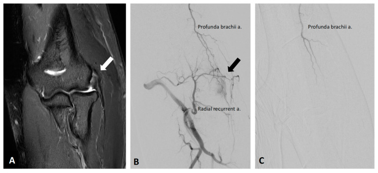
A 58-year-old man with right lateral epicondylitis. A: Preprocedural coronal fat-suppressed T2-weighted MRI shows abnormal thickening and high signal at the common extensor origin (white arrow). B: Preprocedural angiography of the radial recurrent artery demonstrates angiogenesis at the corresponding region, with reflux into the profunda brachii artery (black arrow). C: Postembolization angiography through the profunda brachii and other relevant arteries shows elimination of the abnormal vascularity; pain and function improved after treatment.
Wang B, Liang K-W, Chen C-H, Wang C-K. Diagnostics. 2023;13:134. Original Figure 4. © 2022 by the authors. CC BY 4.0. Image unchanged; caption translated and adapted from the original legend. Read the original full text3|Golfer’s elbow: promising early results, but less evidence than for the lateral side
Lee and colleagues reported a 2022 single-center retrospective cohort of ten patients who underwent 14 procedures for chronic medial epicondylitis after at least three months of conservative treatment. IPM/CS was used in 12 procedures and quick-soluble gelatin sponge in two. Clinical success, defined as more than a 70% reduction in QuickDASH at six months, was achieved after 12 of 14 procedures (85.7%). Mean VAS decreased from 7.6 to 0.9 and mean QuickDASH from 71.9 to 8.4 at six months. No major complication was observed, and improvement persisted in nine patients through follow-up of up to 12 months.
Okuno and Shibuya subsequently discussed the possibility of embolic treatment for medial epicondylitis in a dedicated commentary. Nevertheless, both the number of studies and the total sample size remain substantially smaller for the medial than the lateral elbow, and this difference should be explained during clinical decision-making.
4|MRI, DCE-MRA, and DSA: connecting the lesion to its arterial supply
The 2024 series by Pan, Wang, and colleagues included a 60-year-old man with combined lateral and medial epicondylitis, two years of pain, and a baseline NRS of 9 despite analgesics, rehabilitation, acupuncture, local corticosteroid injection, and platelet-rich plasma injection. MRI demonstrated abnormalities of the common extensor tendon, radial collateral ligament, and common flexor tendon. DCE-MRA depicted angiogenesis on both sides of the elbow and helped identify the inferior ulnar collateral artery (IUCA), posterior ulnar recurrent artery (PURA), radial recurrent artery (RRA), and deep brachial artery (DBA). DSA confirmed the abnormal vascularity and arterial distribution before TAME. The NRS decreased to 2 at six months, with no recorded complication.
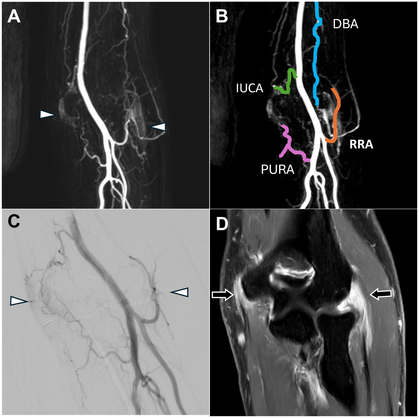
Original Figure 3 combines contrast-enhanced MRI, DCE-MRA, an annotated arterial map, and DSA. MRI demonstrates abnormalities at the medial common flexor and lateral common extensor origins. DCE-MRA and DSA show angiogenesis on both sides of the elbow, while the arterial map labels the IUCA, PURA, RRA, and DBA for preprocedural planning and intraprocedural search.
Pan C-Y, Liang K-W, Chen T-R, et al., Wang B. Front Med (Lausanne). 2024;11:1471504. Original Figure 3. © 2024 Pan et al. CC BY 4.0. Image unchanged; caption adapted from the article and the displayed panel arrangement. The case description reports a left elbow, whereas the original Figure 3 legend reports a right elbow; this page therefore uses “affected elbow” without adjudicating laterality. Read the original full text5|Who should be evaluated? Do not mistake a structural problem for inflammation
When MRI shows a severe CET tear, LUCL injury, radiocapitellar incongruity, UCL tearing, or valgus instability, the structural problem should not be ignored merely because enhancement is also present. See the related articles on MRI of lateral elbow pain and MRI of medial elbow pain.
6|How should the evidence be positioned today?
Published elbow TAME studies suggest possible improvement in pain, function, and selected MRI measures, with few major complications reported in these cohorts. Important limitations remain: small samples, single-center designs, absence of randomized or sham controls, heterogeneity in embolic materials and technique, and differences in patient selection and rehabilitation. Lateral epicondylitis has prospective follow-up to two years; medial epicondylitis is supported mainly by a small retrospective cohort. The most accurate conclusion is not that TAME cures every tennis or golfer’s elbow, but that it is a minimally invasive option worth specialist evaluation for carefully selected patients with a secure diagnosis and persistent symptoms after conservative care.
Take-home messages
- Yuji Okuno and colleagues pioneered musculoskeletal pain embolization and elbow TAME: the 2013 study established the concept, and the 2017 two-year lateral epicondylitis study became a key milestone.
- TAME targets abnormal neovascularity associated with pain; it does not directly repair a tendon or ligament.
- The evidence base is larger for lateral than medial epicondylitis, but both still lack large randomized controlled trials.
- MRI confirms pathology and screens for structural problems; DCE-MRA maps angiogenesis and arterial supply; DSA confirms the intraprocedural target.
- The IUCA, PURA, RRA, and DBA are useful anatomic guides, but embolization targets must be individualized.
- Published results are encouraging, while uncertainty, alternatives, and potential risks still belong in shared decision-making.
Core references and image licensing
1. Okuno Y, Matsumura N, Oguro S. Transcatheter arterial embolization using imipenem/cilastatin sodium for tendinopathy and enthesopathy refractory to nonsurgical management. J Vasc Interv Radiol. 2013;24:787–792. doi:10.1016/j.jvir.2013.02.033.
2. Iwamoto W, Okuno Y, Matsumura N, Kaneko T, Ikegami H. Transcatheter arterial embolization of abnormal vessels as a treatment for lateral epicondylitis refractory to conservative treatment: a pilot study with a 2-year follow-up. J Shoulder Elbow Surg. 2017;26:1335–1341. doi:10.1016/j.jse.2017.03.026.
3. Hwang JH, Park SW, Kim KH, et al. Early Results of Transcatheter Arterial Embolization for Relief of Chronic Shoulder or Elbow Pain Associated with Tendinopathy Refractory to Conservative Treatment. J Vasc Interv Radiol. 2018;29:510–517. doi:10.1016/j.jvir.2017.11.013.
4. Lee JH, Kim DH, Lee SH, et al. Short-term Results of Transcatheter Arterial Embolization for Chronic Medial Epicondylitis Refractory to Conservative Treatment. Cardiovasc Intervent Radiol. 2022;45:197–204. doi:10.1007/s00270-021-02878-2. See also: Okuno Y, Shibuya M. Possibility of Embolic Treatment for Medial Epicondylitis. doi:10.1007/s00270-021-03008-8.
5. Wang B, Liang K-W, Chen C-H, Wang C-K. Transcatheter Arterial Embolization for Alleviating Chronic Musculoskeletal Pain and Improving Physical Function: A Narrative Review. Diagnostics. 2023;13:134. doi:10.3390/diagnostics13010134. Figure 4: © 2022 by the authors, CC BY 4.0.
6. Pan C-Y, Liang K-W, Chen T-R, et al., Wang B. Added value of pre-procedural magnetic resonance angiography in transarterial embolization for refractory musculoskeletal pain. Front Med (Lausanne). 2024;11:1471504. doi:10.3389/fmed.2024.1471504. Figure 3: © 2024 Pan et al., CC BY 4.0.
This page reproduces only Figure 4 and Figure 3 from the two CC BY 4.0 articles above. The non-CC BY papers by Okuno, Iwamoto, Hwang, and Lee are cited for scholarship; their figures are not reproduced.
This page is for medical education and academic communication. It does not replace individual diagnosis or treatment advice.
Continue reading this category →