
Musculoskeletal Imaging Education
MRI of Medial Elbow Pain: From Golfer’s Elbow to Valgus Instability
Medial elbow pain often suggests medial epicondylitis or golfer’s elbow. Common flexor tendinopathy, medial collateral ligament injury, and ulnar neuropathy can produce overlapping symptoms and may coexist. A useful MRI approach connects the superficial tendon, deeper ligament, osseous attachments, and cubital tunnel.
1|Know the normal medial stabilizers: anterior and posterior bundles
The medial collateral ligament (MCL), also called the ulnar collateral ligament (UCL), has anterior, posterior, and transverse bundles. The anterior bundle (A-MCL) runs from the inferior medial epicondyle to the sublime tubercle of the ulnar coronoid process. Its anterior band is an important static restraint to valgus and internal rotation; the anterior bundle is the primary medial stabilizer at approximately 30–120° of elbow flexion. The posterior bundle (P-MCL) runs from the posterior medial epicondyle to the medial olecranon, forms the cubital-tunnel floor, and contributes secondary stability beyond 90° of flexion. The transverse bundle has both attachments on the ulna and contributes little stability.
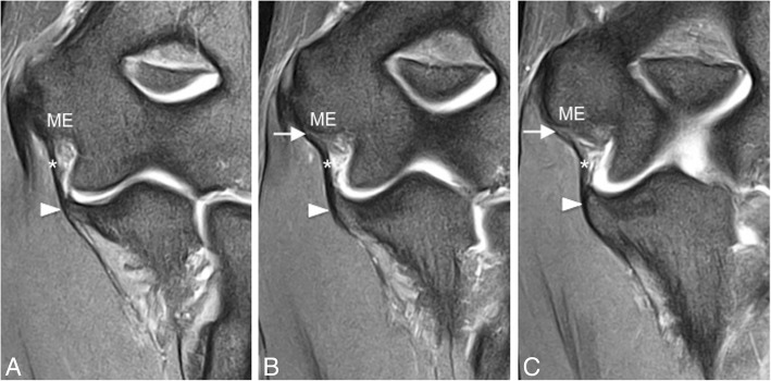
Sequential coronal fat-suppressed proton-density-weighted MRI (a–c) demonstrates an intact anterior bundle of the MCL (A-MCL; white asterisks). It runs from the inferior medial epicondyle (ME) to the sublime tubercle of the ulna (white arrowheads). White arrows identify the typical striated appearance of the normal proximal A-MCL.
Original Fig. 5. Acosta Batlle J, Cerezal L, López Parra MD, Alba B, Resano S, Blázquez Sánchez J. The elbow: review of anatomy and common collateral ligament complex pathology using MRI. Insights Imaging. 2019;10:43. doi:10.1186/s13244-019-0725-7. © The Author(s) 2019. CC BY 4.0. Caption adapted from original legend.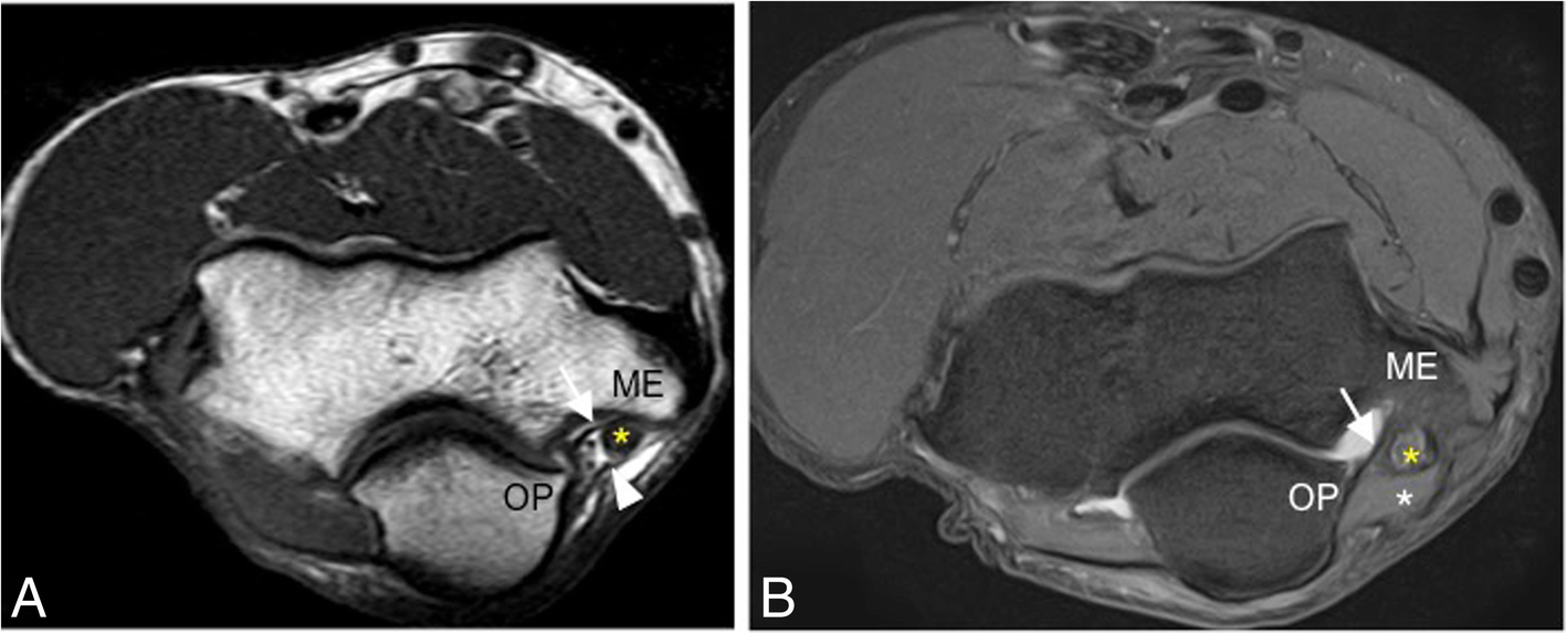
Axial T1-weighted MRI (a) and axial fat-suppressed proton-density-weighted MRI (b), from two different healthy volunteers, show an intact posterior MCL bundle (white arrows). It extends from the posterior medial epicondyle (ME) to the medial olecranon process (OP). The white arrowhead marks the cubital tunnel retinaculum; yellow asterisks mark the ulnar nerve, and the white asterisk marks the anconeus epitrochlearis muscle.
Original Fig. 6. Acosta Batlle J, Cerezal L, López Parra MD, Alba B, Resano S, Blázquez Sánchez J. The elbow: review of anatomy and common collateral ligament complex pathology using MRI. Insights Imaging. 2019;10:43. doi:10.1186/s13244-019-0725-7. © The Author(s) 2019. CC BY 4.0. Caption adapted from original legend.2|Golfer’s elbow (medial epicondylitis): focus on the common flexor tendon
Golfer’s elbow is the common name for medial epicondylitis. MRI evaluation centers on the common flexor tendon (CFT) origin. Original Fig. 10 demonstrates diffuse tendon thickening, intermediate intratendinous signal, and adjacent muscle edema across three planes. Assess thickness, signal, and fiber continuity, then review adjacent muscles and the deeper UCL. Altered signal does not itself establish tearing: the original legend describes tendinosis, not a partial- or full-thickness tear.
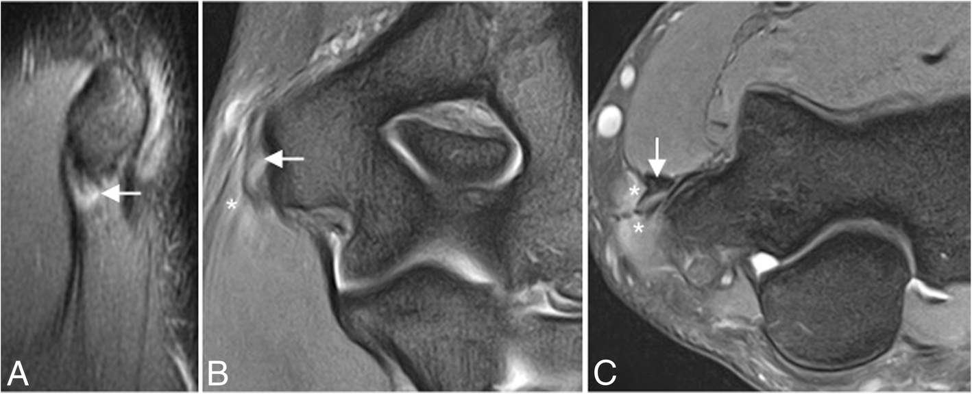
A 45-year-old man with medial elbow pain. Sagittal proton-density-weighted MRI (a), coronal fat-suppressed proton-density-weighted MRI (b), and axial fat-suppressed proton-density-weighted MRI (c) show common flexor tendinosis: intermediate signal within a diffusely thickened flexor tendon origin (white arrows). White asterisks mark edema in the flexor digitorum superficialis and palmaris longus muscles.
Original Fig. 10. Acosta Batlle J, Cerezal L, López Parra MD, Alba B, Resano S, Blázquez Sánchez J. The elbow: review of anatomy and common collateral ligament complex pathology using MRI. Insights Imaging. 2019;10:43. doi:10.1186/s13244-019-0725-7. © The Author(s) 2019. CC BY 4.0. Caption adapted from original legend.3|Acute anterior-bundle injury: inspect both attachments and the midsubstance
After a fall on an outstretched arm, assess for anterior-bundle tearing, attachment avulsion, and associated contusions. Describe the location, partial- or full-thickness disruption, fluid gap, and any osseous fragment. Continue tracing the entire ligament after finding one injury. Original Figs. 14 and 15 are acute post-fall cases illustrating injury combinations associated with medial instability; they are not chronic throwing-overuse cases.
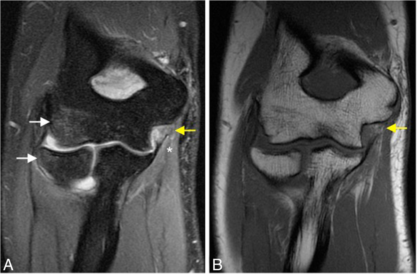
A 23-year-old man with pain and medial instability after a fall on an outstretched arm. Coronal fat-suppressed proton-density-weighted MRI (a) and coronal T1-weighted MRI (b) show an acute full-thickness tear of the proximal anterior MCL bundle (yellow arrows), FDS muscle edema (white asterisk), radial-head and posterior capitellar contusions (white arrows), and joint effusion.
Original Fig. 14. Acosta Batlle J, Cerezal L, López Parra MD, Alba B, Resano S, Blázquez Sánchez J. The elbow: review of anatomy and common collateral ligament complex pathology using MRI. Insights Imaging. 2019;10:43. doi:10.1186/s13244-019-0725-7. © The Author(s) 2019. CC BY 4.0. Caption adapted from original legend.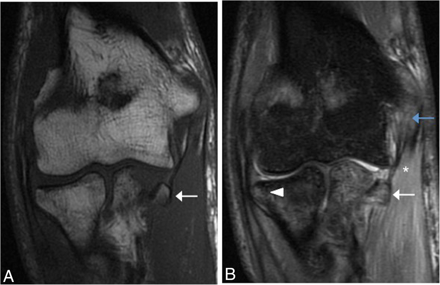
A 32-year-old man with pain and medial instability after a fall on an outstretched arm. Coronal T1-weighted MRI (a) and coronal fat-suppressed proton-density-weighted MRI (b) show an acute avulsion fracture at the anterior MCL bundle insertion on the sublime tubercle (white arrows), an acute proximal partial-thickness tear of the same bundle (blue arrow), FDS edema (white asterisk), and radial-head contusion (white arrowhead).
Original Fig. 15. Acosta Batlle J, Cerezal L, López Parra MD, Alba B, Resano S, Blázquez Sánchez J. The elbow: review of anatomy and common collateral ligament complex pathology using MRI. Insights Imaging. 2019;10:43. doi:10.1186/s13244-019-0725-7. © The Author(s) 2019. CC BY 4.0. Caption adapted from original legend.4|Chronic valgus loading: a thick ligament may still be abnormal
Repeated valgus stress produces medial tension, lateral compression, and extension overload. Chronic MCL injury may appear thickened, show abnormal signal, or lose continuity. Inspect the sublime tubercle for deformity from an old fracture. MRI characterizes structural changes; functional valgus instability requires correlation with symptoms, history, and clinical stability assessment.
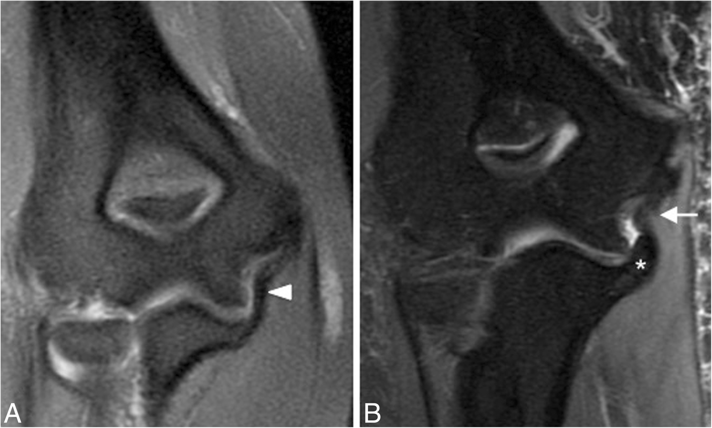
Two different patients with chronic elbow pain and medial instability. Coronal fat-suppressed proton-density-weighted MRI (a) shows chronic thickening of the anterior MCL bundle (white arrowhead). In the second patient, the same coronal sequence (b) shows chronic anterior-bundle thickening (white arrow) and deformity of the sublime tubercle from malunion of an old fracture (white asterisk).
Original Fig. 18. Acosta Batlle J, Cerezal L, López Parra MD, Alba B, Resano S, Blázquez Sánchez J. The elbow: review of anatomy and common collateral ligament complex pathology using MRI. Insights Imaging. 2019;10:43. doi:10.1186/s13244-019-0725-7. © The Author(s) 2019. CC BY 4.0. Caption adapted from original legend.5|Posterior bundle and ulnar nerve: include the cubital tunnel
Paresthesias, sensory symptoms, or weakness should prompt a targeted review of the ulnar nerve and cubital tunnel. Because the posterior bundle forms the tunnel floor, ligament injury and surrounding soft-tissue changes may accompany altered nerve position and shape. Assess nerve course, caliber, signal, flattening or displacement, and adjacent bone, ligament, and muscle.
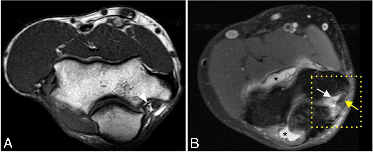
The injured patient is a 25-year-old man with pain and medial instability after a fall on an outstretched hand. Panel a is axial T1-weighted MRI of a healthy volunteer’s right elbow, showing an intact posterior MCL bundle (white arrowhead). Panel b is axial fat-suppressed proton-density-weighted MRI of the patient’s left elbow, showing an acute posterior-bundle tear (white arrow), inflammatory cubital-tunnel soft-tissue changes (white asterisk), displacement and flattening of the ulnar nerve (yellow arrow), and joint effusion (black asterisks).
Original Fig. 17. Acosta Batlle J, Cerezal L, López Parra MD, Alba B, Resano S, Blázquez Sánchez J. The elbow: review of anatomy and common collateral ligament complex pathology using MRI. Insights Imaging. 2019;10:43. doi:10.1186/s13244-019-0725-7. © The Author(s) 2019. CC BY 4.0. Caption adapted from original legend.6|Young throwers: inspect the medial epicondylar physis
In the immature elbow, repeated traction by the CFT and MCL can injure the medial epicondylar apophysis, producing findings associated with Little Leaguer’s elbow. Assess physeal width, marrow edema, and possible avulsion in addition to ligament integrity. Do not apply an adult tendon-and-ligament pattern without considering skeletal maturity.
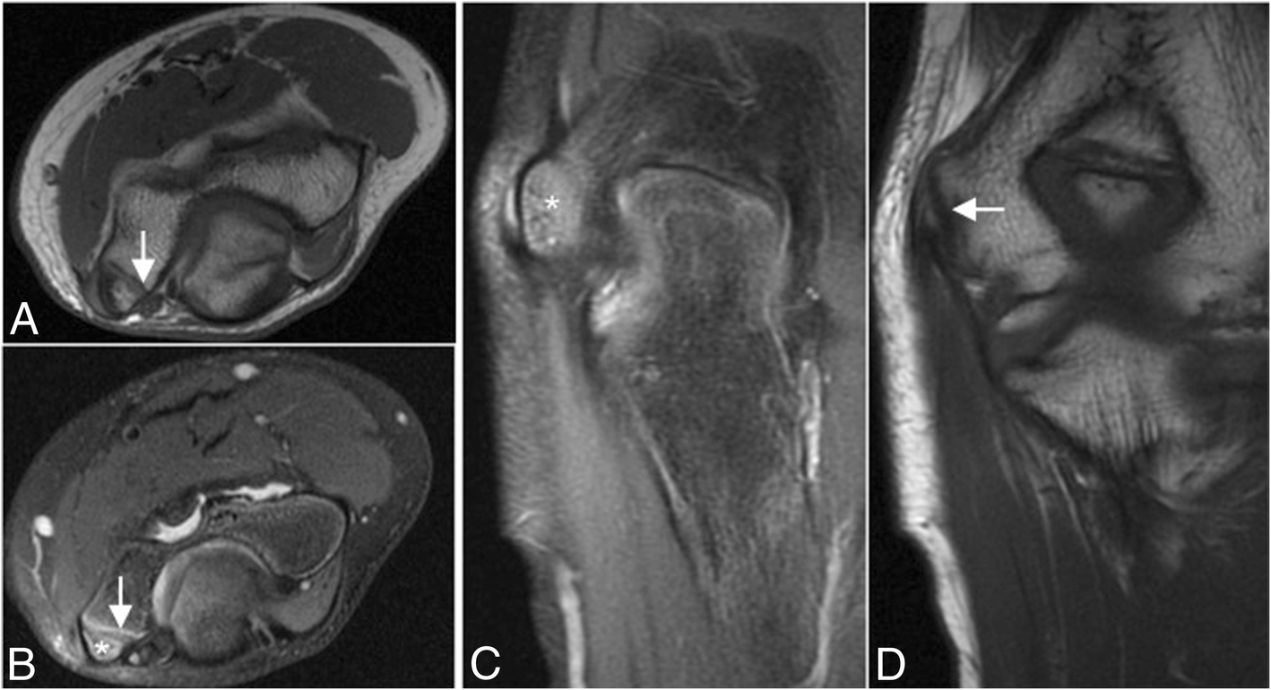
A 12-year-old left-handed baseball pitcher with medial epicondylar pain. Axial T1-weighted MRI (a), axial fat-suppressed proton-density-weighted MRI (b), coronal fat-suppressed proton-density-weighted MRI (c), and coronal T1-weighted MRI (d) show widening of the medial epicondylar physis (white arrows) and associated bone marrow edema (white asterisks).
Original Fig. 20. Acosta Batlle J, Cerezal L, López Parra MD, Alba B, Resano S, Blázquez Sánchez J. The elbow: review of anatomy and common collateral ligament complex pathology using MRI. Insights Imaging. 2019;10:43. doi:10.1186/s13244-019-0725-7. © The Author(s) 2019. CC BY 4.0. Caption adapted from original legend.Practical MRI Checklist
Take-home Messages
- Medial elbow pain is not synonymous with golfer’s elbow: review tendon, ligament, nerve, and physis.
- Normal proximal anterior-bundle striation should not be labeled a tear by itself.
- Trace the anterior bundle end to end: more than one injury site can coexist.
- Muscle edema, contusions, and effusion provide clues but do not independently establish valgus instability.
- Paresthesias or weakness warrant evaluation of the posterior bundle, cubital tunnel, and ulnar nerve.
- In young throwers, inspect the medial epicondylar physis; correlate MRI structural findings with clinical stability.
Primary source and image license
Acosta Batlle J, Cerezal L, López Parra MD, Alba B, Resano S, Blázquez Sánchez J. The elbow: review of anatomy and common collateral ligament complex pathology using MRI. Insights Imaging. 2019;10:43. doi:10.1186/s13244-019-0725-7.
© The Author(s) 2019. Creative Commons Attribution 4.0 International License (CC BY 4.0).
Original Figures 5, 6, 10, 14, 15, 18, 17, and 20 are reproduced with all panels and markers retained; no cropping or new image annotations. Captions are translated/adapted from the original legends. Teaching points and the reading checklist are an educational synthesis, separate from the original legends.
This page is for medical education and academic communication. It does not replace individual diagnosis or treatment advice.
Continue reading this category →