
Musculoskeletal Imaging Education
MRI of Lateral Elbow Pain: Beyond Tennis Elbow
Lateral elbow pain often leads directly to lateral epicondylitis or common extensor tendinopathy. However, stopping once an abnormal common extensor tendon (CET) is identified may miss clinically important lateral collateral ligament complex injury and instability.
1|Start with normal lateral elbow anatomy
The lateral collateral ligament complex consists primarily of the radial collateral ligament (RCL), lateral ulnar collateral ligament (LUCL), and annular ligament. The RCL lies deep to the CET. The LUCL is a key stabilizer against posterolateral rotatory instability (PLRI), while the common extensor origin also contributes secondary soft-tissue stability.
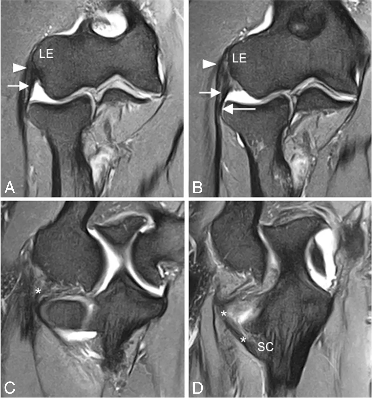
Sequential coronal fat-suppressed proton-density-weighted MR images (a–d) show an intact lateral ulnar collateral ligament (LUCL; white asterisks) and radial collateral ligament (RCL; short arrows). The LUCL courses around the posterior aspect of the radial neck. The annular ligament is marked by the long arrow, and the common extensor tendon by white arrowheads. LE = lateral epicondyle; SC = supinator crest of the ulna.
Acosta Batlle J, et al. Insights Imaging. 2019;10:43. Original Fig. 7. CC BY 4.0. Caption adapted from the original figure legend.2|CET: tendinosis → partial tear → full-thickness tear
Common MRI findings include tendon thickening, increased intratendinous signal, and architectural distortion. The key principle is that increased signal does not necessarily equal a tear; first assess tendon fiber continuity.
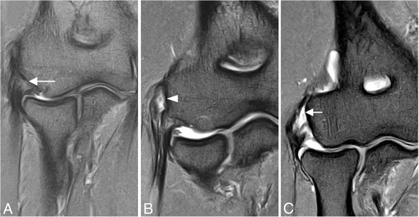
Three different patients with lateral elbow pain. Coronal fat-suppressed proton-density-weighted MR images (a–c) show common extensor tendinosis (long arrow), a partial-thickness common extensor tendon tear (arrowhead), and a full-thickness common extensor tendon tear (short arrow), respectively.
Acosta Batlle J, et al. Insights Imaging. 2019;10:43. Original Fig. 9. CC BY 4.0. Caption adapted from the original figure legend.3|Don’t stop at the tendon
Advanced lateral epicondylitis is not necessarily isolated tendinopathy. With severe CET pathology, continue to assess the RCL, LUCL, radiocapitellar congruity, and radial-head or capitellar bone contusions. Clicking, snapping, locking, giving-way, previous dislocation, or prior lateral elbow surgery should further raise concern for instability.
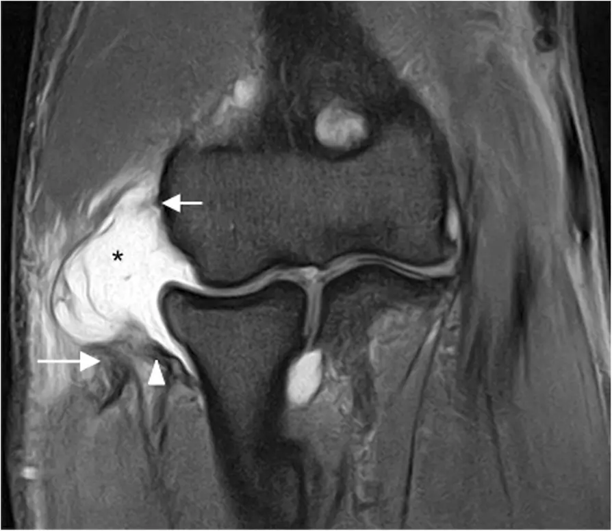
Stage 2 posterolateral rotatory instability. Coronal fat-suppressed proton-density-weighted MRI shows complete detachment of the proximal LUCL, proximal RCL proper, and common extensor tendon (short white arrow), with retraction of the LUCL (white arrowhead) and CET (long white arrow). Joint fluid extravasates laterally through the tear (black asterisk).
Acosta Batlle J, et al. Insights Imaging. 2019;10:43. Original Fig. 24. CC BY 4.0. Caption adapted from the original figure legend.4|LUCL injury and PLRI
Evaluate the LUCL on coronal oblique, coronal, and axial images. Sagittal images are particularly useful for detecting associated posterolateral radial-head subluxation.
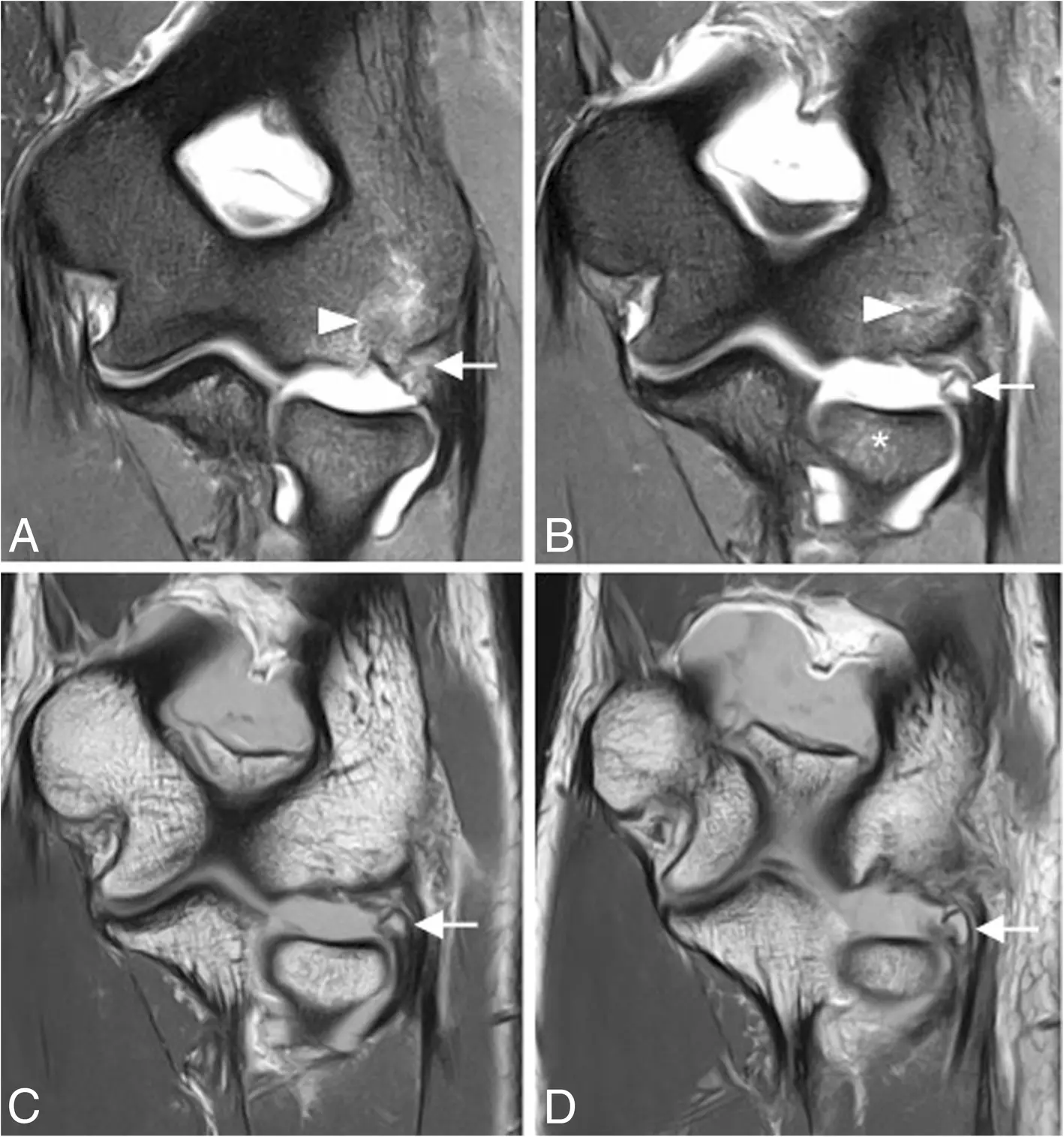
Stage 1 posterolateral rotatory instability. Coronal fat-suppressed proton-density-weighted MR images (a, b) and coronal T1-weighted MR images (c, d) show proximal LUCL avulsion with retraction of an osseous fragment (white arrows). Bone marrow edema is present in the capitellum (white arrowheads) and radial head (white asterisks).
Acosta Batlle J, et al. Insights Imaging. 2019;10:43. Original Fig. 23. CC BY 4.0. Caption adapted from the original figure legend.5|Bone contusions can reveal the injury mechanism
Do not rely only on a structure-by-structure checklist. Posterolateral capitellar or radial-head contusions, joint incongruity, and radial-head translation can collectively provide clues to the mechanism of instability.
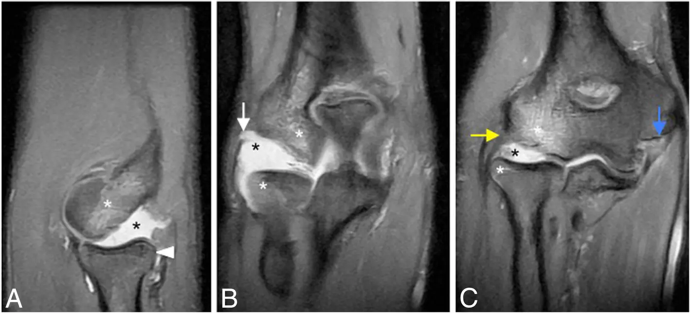
Stage 3B posterolateral rotatory instability. Sagittal fat-suppressed proton-density-weighted MRI (a) and sequential coronal fat-suppressed proton-density-weighted MR images (b, c) show posterolateral radial-head subluxation (white arrowhead), proximal LUCL disruption (white arrow), partial proximal RCL disruption (yellow arrow), and disruption of the proximal anterior bundle of the medial collateral ligament (blue arrow). Radial-head and posterior capitellar contusions (white asterisks) and joint effusion (black asterisks) are also present.
Acosta Batlle J, et al. Insights Imaging. 2019;10:43. Original Fig. 30. CC BY 4.0. Caption adapted from the original figure legend.6|Pain plus clicking: think beyond the tendon
When lateral elbow pain is accompanied by clicking, snapping, or locking, broaden the differential diagnosis to include PLRI, annular-ligament pathology, posterolateral plica, loose bodies, and osteochondral lesions.
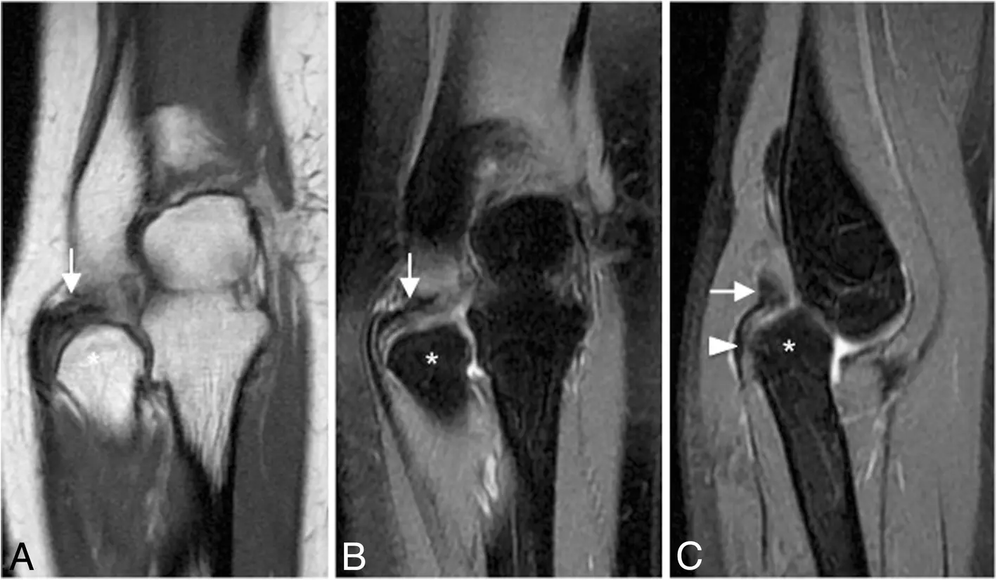
A 40-year-old man with recurrent painful clicking. Coronal T1-weighted MRI (a), coronal fat-suppressed proton-density-weighted MRI (b), and sagittal fat-suppressed proton-density-weighted MRI (c) show chronic annular ligament rupture and displacement (white arrows), escape of the radial head from the annular-ligament sling (white arrowhead), and associated radial-head deformity (white asterisks).
Acosta Batlle J, et al. Insights Imaging. 2019;10:43. Original Fig. 33. CC BY 4.0. Caption adapted from the original figure legend.Practical MRI checklist
Take-home messages
- Lateral elbow pain is not synonymous with lateral epicondylitis.
- Severe CET pathology should trigger careful assessment of the deeper RCL and LUCL.
- LUCL injury may be the missing diagnosis in “refractory tennis elbow.”
- Pain with clicking or locking should broaden the differential diagnosis.
- MRI should identify not only the lesion but also the injury mechanism.
Primary source and image license
Acosta Batlle J, Cerezal L, López Parra MD, et al. The elbow: review of anatomy and common collateral ligament complex pathology using MRI. Insights Imaging. 2019;10:43. doi:10.1186/s13244-019-0725-7.
The original article and figures are distributed under the Creative Commons Attribution 4.0 International License (CC BY 4.0).
This page is for medical education and academic communication. It does not replace individual diagnosis or treatment advice.
Continue reading this category →