
外側肘痛最常讓人想到外側上髁炎/伸肌總腱病變(lateral epicondylitis/common extensor tendinopathy);但磁振造影(MRI)若看到伸肌總腱(common extensor tendon, CET)異常就停止判讀,可能漏掉外側副韌帶複合體病變(lateral collateral ligament complex pathology)與不穩定(instability)。
1|先認識正常的外側肘解剖(lateral elbow anatomy)
外側韌帶複合體主要包含橈側副韌帶(radial collateral ligament, RCL)、外側尺側副韌帶(lateral ulnar collateral ligament, LUCL)與環狀韌帶(annular ligament)。橈側副韌帶位於伸肌總腱深部;外側尺側副韌帶是抵抗後外側旋轉不穩定(posterolateral rotatory instability, PLRI)的重要穩定結構(stabilizer),而伸肌總腱起點(CET origin)也是外側肘的次要軟組織穩定結構(secondary soft-tissue stabilizer)。
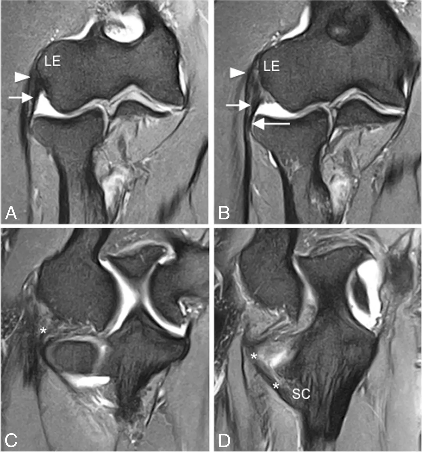
連續冠狀面脂肪抑制質子密度加權磁振造影(coronal FS PD-weighted MRI;a–d)顯示完整的外側尺側副韌帶(lateral ulnar collateral ligament, LUCL;白色星號)與橈側副韌帶(radial collateral ligament, RCL;短箭頭)。外側尺側副韌帶繞過橈骨頸後側(posterior aspect of the radial neck);環狀韌帶(annular ligament)以長箭頭標示,伸肌總腱(common extensor tendon)以白色箭頭標示。LE 代表外上髁(lateral epicondyle);SC 代表尺骨旋後肌嵴(supinator crest of the ulna)。
Acosta Batlle J, et al. Insights Imaging. 2019;10:43. Original Fig. 7. CC BY 4.0. Caption translated and adapted from the original figure legend.2|伸肌總腱(CET):腱病變(tendinosis)→ 部分厚度撕裂(partial-thickness tear)→ 全層撕裂(full-thickness tear)
磁振造影(MRI)常見肌腱增厚(tendon thickening)、腱內訊號升高(increased intratendinous signal)與纖維結構改變。重點是:訊號升高(increased signal)≠ 撕裂(tear);應先確認肌腱纖維的連續性(tendon fiber continuity)。
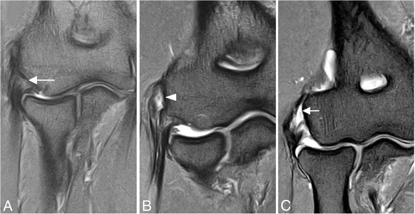
三位不同的外側肘痛(lateral elbow pain)病人。冠狀面脂肪抑制質子密度加權磁振造影(coronal FS PD-weighted MRI;a–c)依序顯示伸肌總腱病變(common extensor tendinosis;長箭頭)、伸肌總腱部分厚度撕裂(common extensor tendon partial-thickness tear;箭頭),以及伸肌總腱全層撕裂(common extensor tendon full-thickness tear;短箭頭)。
Acosta Batlle J, et al. Insights Imaging. 2019;10:43. Original Fig. 9. CC BY 4.0. Caption translated and adapted from the original figure legend.3|不要只看到肌腱就停止判讀(Don’t stop at the tendon)
嚴重外側上髁炎(advanced lateral epicondylitis)不一定只是腱病變(tendinopathy)。看到嚴重伸肌總腱病變(severe CET pathology)時,應繼續確認橈側副韌帶(RCL)、外側尺側副韌帶(LUCL)、肱橈關節對位(radiocapitellar congruity),以及橈骨頭/肱骨小頭骨挫傷(radial head/capitellar bone contusion)。若合併彈響(clicking/snapping)、卡住(locking)、無力感(giving-way)、舊脫臼或外側肘手術史(lateral elbow surgery history),更應提高對不穩定(instability)的警覺。
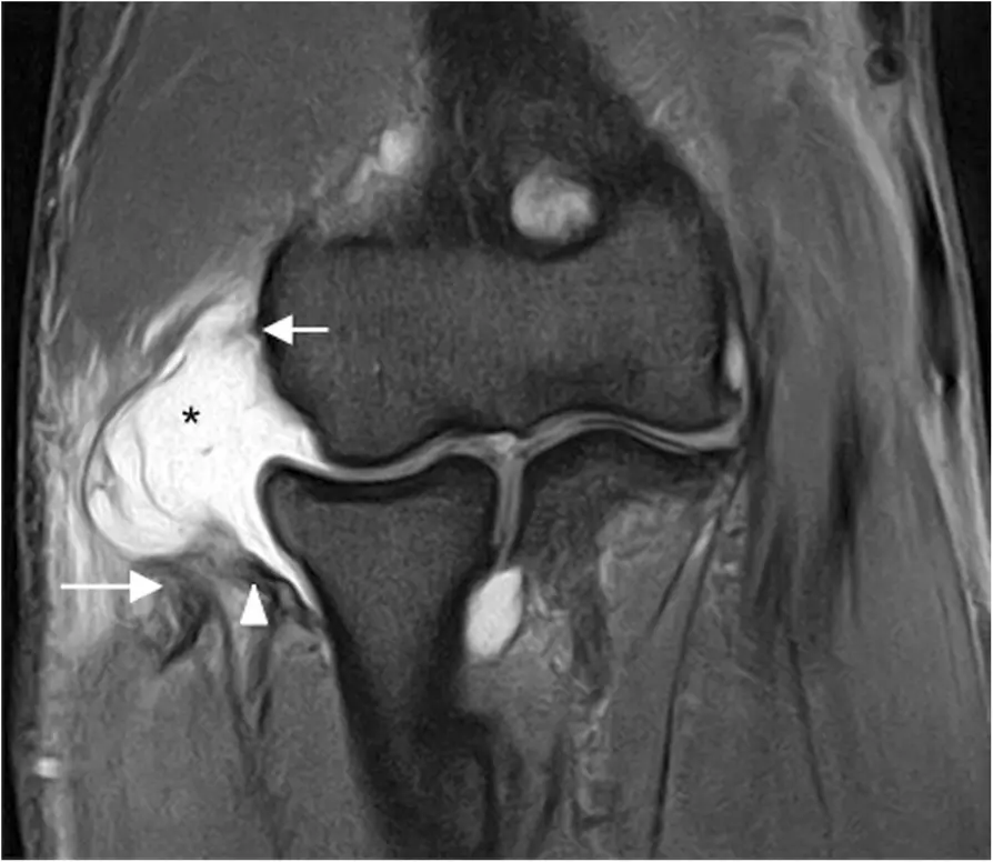
後外側旋轉不穩定(posterolateral rotatory instability, PLRI)第 2 期。冠狀面脂肪抑制質子密度加權磁振造影(coronal FS PD-weighted MRI)顯示近端外側尺側副韌帶(proximal LUCL)、近端橈側副韌帶本體(proximal RCL proper)與伸肌總腱(common extensor tendon)完全撕脫(detachment;白色短箭頭);外側尺側副韌帶(LUCL;白色箭頭)及伸肌總腱(CET;白色長箭頭)均有回縮(retraction)。另可見關節液(joint fluid)經撕裂處向外滲出(extravasation;黑色星號)。
Acosta Batlle J, et al. Insights Imaging. 2019;10:43. Original Fig. 24. CC BY 4.0. Caption translated and adapted from the original figure legend.4|外側尺側副韌帶損傷(LUCL injury)與後外側旋轉不穩定(PLRI)
外側尺側副韌帶損傷(LUCL injury)建議在斜冠狀面、冠狀面與軸位影像(coronal oblique, coronal and axial planes)評估;相關的橈骨頭後外側半脫位(associated posterolateral radial head subluxation)則以矢狀面影像(sagittal images)的評估特別重要。
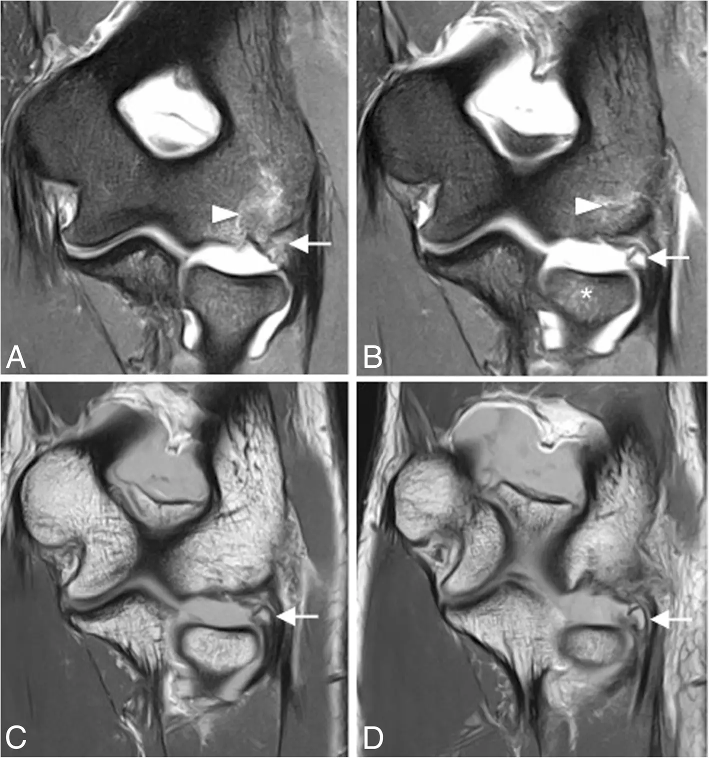
後外側旋轉不穩定(posterolateral rotatory instability, PLRI)第 1 期。冠狀面脂肪抑制質子密度加權磁振造影(coronal FS PD-weighted MRI;a, b)與冠狀面 T1 加權磁振造影(coronal T1-weighted MRI;c, d)顯示近端外側尺側副韌帶撕脫(proximal LUCL avulsion),並有骨片回縮(osseous fragment retraction;白色箭頭)。肱骨小頭(capitellum)可見骨髓水腫(bone marrow edema;白色箭頭),橈骨頭(radial head)也有骨髓水腫(白色星號)。
Acosta Batlle J, et al. Insights Imaging. 2019;10:43. Original Fig. 23. CC BY 4.0. Caption translated and adapted from the original figure legend.5|骨挫傷(bone contusion)也在告訴你受傷機轉(injury mechanism)
不要只做逐一結構檢查表(structure-by-structure checklist)。肱骨小頭後外側/橈骨頭骨挫傷(posterolateral capitellar/radial head contusion)、關節對位不良(joint incongruity)與橈骨頭位移(radial head translation),可以共同提供不穩定機轉(instability mechanism)的線索。
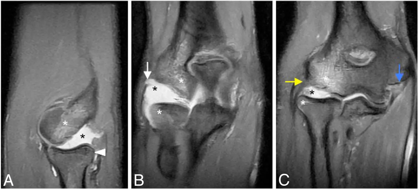
後外側旋轉不穩定(posterolateral rotatory instability, PLRI)第 3B 期。矢狀面脂肪抑制質子密度加權磁振造影(sagittal FS PD-weighted MRI;a)及連續冠狀面脂肪抑制質子密度加權磁振造影(coronal FS PD-weighted MRI;b, c)顯示橈骨頭後外側半脫位(radial head posterolateral subluxation;白色箭頭)、近端外側尺側副韌帶斷裂(proximal LUCL disruption;白色箭頭)、近端橈側副韌帶部分斷裂(proximal partial RCL disruption;黃色箭頭)及內側副韌帶前束近端斷裂(proximal anterior bundle of MCL disruption;藍色箭頭)。另可見橈骨頭與肱骨小頭後方骨挫傷(radial head and posterior capitellar contusions;白色星號),以及關節積液(joint effusion;黑色星號)。
Acosta Batlle J, et al. Insights Imaging. 2019;10:43. Original Fig. 30. CC BY 4.0. Caption translated and adapted from the original figure legend.6|疼痛合併彈響(pain + clicking):不要只想到肌腱(tendon)
當外側肘痛(lateral elbow pain)合併彈響(clicking/snapping)或卡住(locking),鑑別診斷(differential diagnosis)應擴大到後外側旋轉不穩定(PLRI)、環狀韌帶病變(annular ligament pathology)、後外側滑膜皺襞(posterolateral plica)、關節內游離體(loose body)與骨軟骨病灶(osteochondral lesion)等。
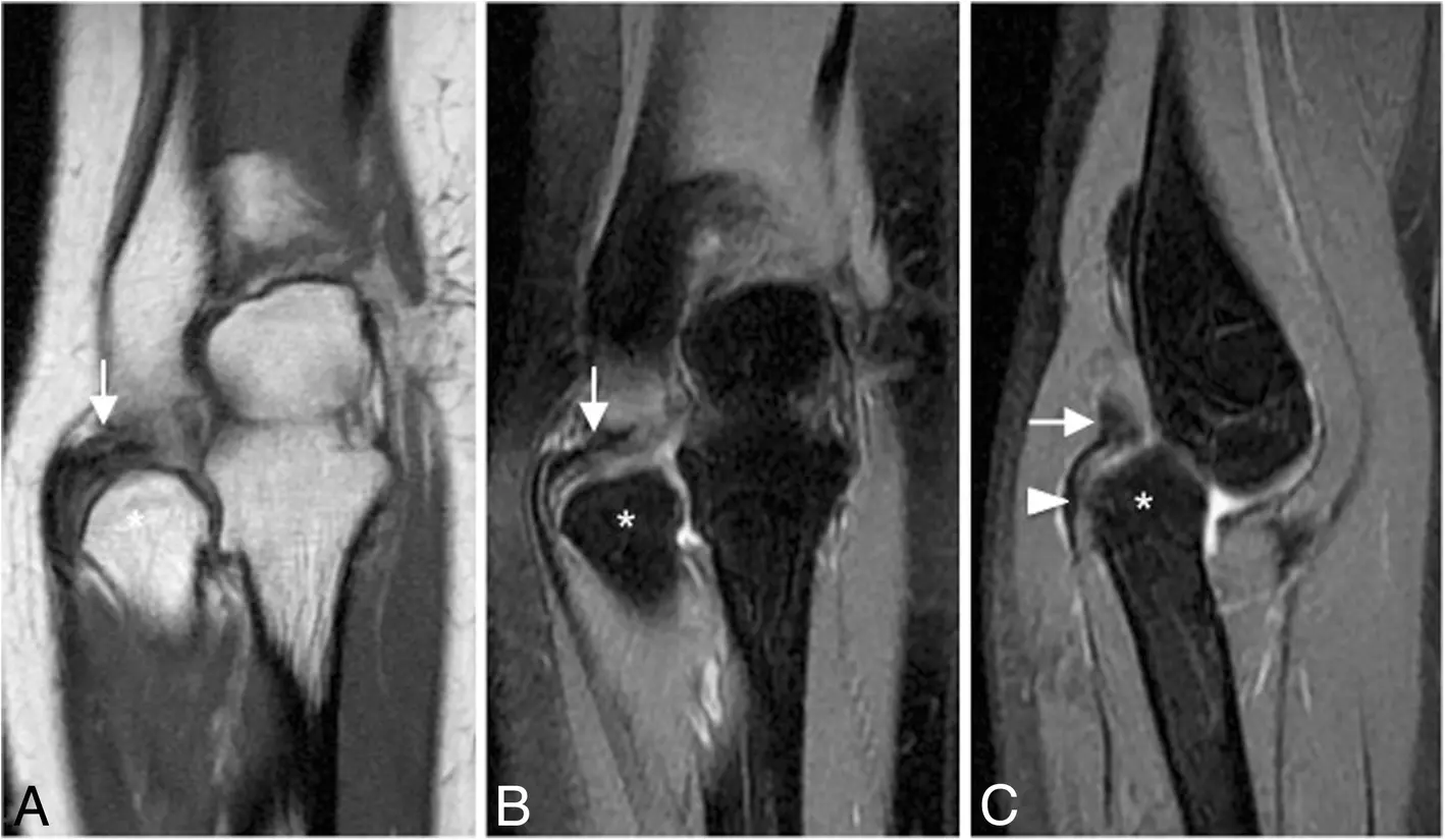
40 歲男性,反覆出現疼痛性彈響(painful clicking)。冠狀面 T1 加權磁振造影(coronal T1-weighted MRI;a)、冠狀面脂肪抑制質子密度加權磁振造影(coronal FS PD-weighted MRI;b)及矢狀面脂肪抑制質子密度加權磁振造影(sagittal FS PD-weighted MRI;c)顯示慢性環狀韌帶斷裂與移位(chronic annular ligament rupture and displacement;白色箭頭)、橈骨頭(radial head)脫出環狀韌帶套環(annular ligament sling;白色箭頭),並伴隨橈骨頭變形(radial head deformity;白色星號)。
Acosta Batlle J, et al. Insights Imaging. 2019;10:43. Original Fig. 33. CC BY 4.0. Caption translated and adapted from the original figure legend.實用磁振造影檢查表(Practical MRI checklist)
重點整理(Take-home messages)
- 外側肘痛(lateral elbow pain)不等於只有外側上髁炎(lateral epicondylitis)。
- 出現嚴重伸肌總腱病變(severe CET pathology)時,一定要往深層檢查橈側副韌帶(RCL)與外側尺側副韌帶(LUCL)。
- 漏掉外側尺側副韌帶損傷(LUCL injury),可能把不穩定(instability)當成頑固性網球肘(refractory tennis elbow)。
- 疼痛合併彈響或卡住(pain + clicking/locking)時,要擴大鑑別診斷(differential diagnosis)。
- 磁振造影(MRI)不只用來找病灶,也要讀懂受傷機轉(injury mechanism)。
主要文獻與圖片授權
Acosta Batlle J, Cerezal L, López Parra MD, et al. The elbow: review of anatomy and common collateral ligament complex pathology using MRI. Insights Imaging. 2019;10:43. doi:10.1186/s13244-019-0725-7.
原文與圖片依 Creative Commons Attribution 4.0 International License(CC BY 4.0)發布。
本文為醫療衛教與學術交流用途,不能取代個別診斷與治療建議。
繼續閱讀此分類文章 →