
Musculoskeletal Imaging Education
Hoffa’s Fat Pad and Anterior Knee Pain: An MRI Reading Pathway
On knee MRI, high signal in Hoffa’s infrapatellar fat pad (HFP) often attracts attention in patients with anterior knee pain. Edema is a starting point, not a diagnosis: it may accompany impingement, trauma, postoperative change or adjacent disease, and can also be asymptomatic. Using the 2016 pictorial review by Draghi and colleagues, this article follows anatomy, signal distribution and the source of pathology rather than attributing all anterior knee pain to the fat pad.
1|Start with normal anatomy: fat, recesses and plicae
The HFP is intracapsular and extrasynovial, posterior to the patellar tendon and anterior to the synovium, with attachments to the anterior meniscal horns. Normal fat is bright on non-fat-suppressed T1- and T2-weighted images, while fibrous septa are dark. Vertical suprahoffatic and horizontal infrahoffatic recesses may contain joint fluid; their normal configuration should be recognized before diagnosing a cyst or a tear.
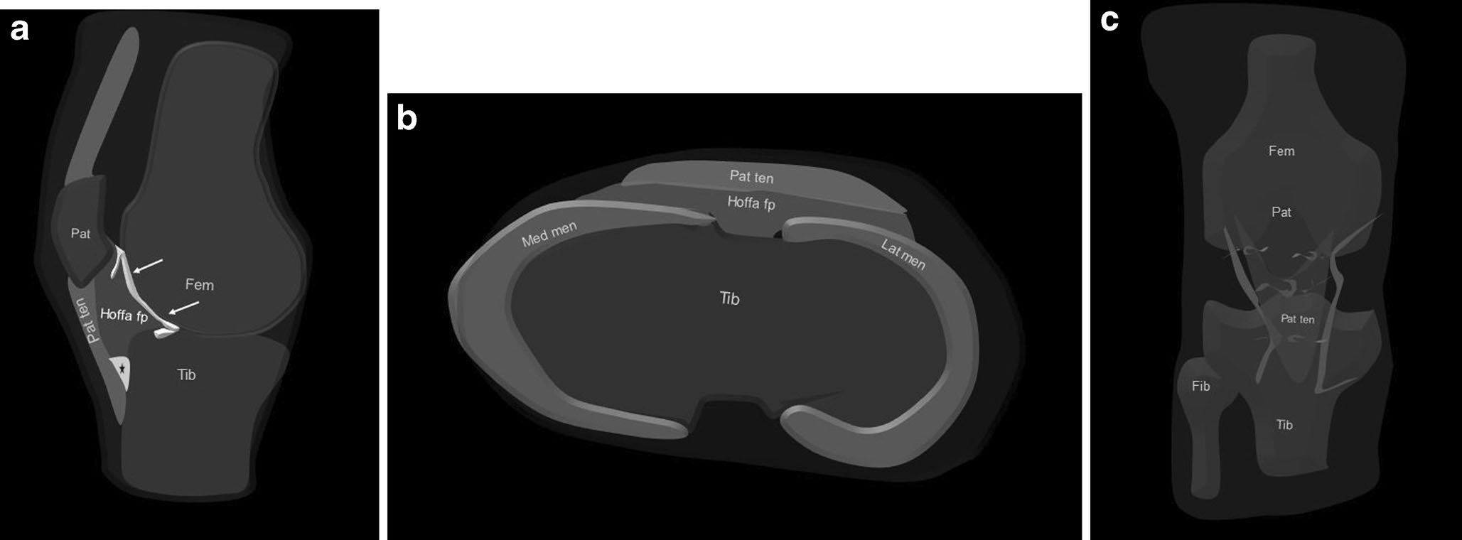
Anatomical diagrams, not MR images. a: The HFP (Hoffa fp) is bounded anteriorly by the patellar tendon (Pat ten) and capsule, superiorly by the lower patellar pole (Pat), inferiorly by the proximal tibia (Tib) and deep infrapatellar bursa (asterisk), and posteriorly by the synovium (arrows) and femur (Fem). b: It attaches directly to the anterior horns of the medial and lateral menisci (Med men, Lat men). c: Two vertical arteries run posterior and parallel to the lateral edges of the patellar tendon.
Display adaptation: panel b is vertically scaled to match the height of panels a and c. The original is available via the Original Fig. 1 link.
Original Fig. 1. Draghi F, Ferrozzi G, Urciuoli L, Bortolotto C, Bianchi S. Hoffa’s fat pad abnormalities, knee pain and magnetic resonance imaging in daily practice. Insights Imaging. 2016;7(3):373–383. doi:10.1007/s13244-016-0483-8. © The Author(s) 2016. CC BY 4.0. Caption translated/adapted from original legend.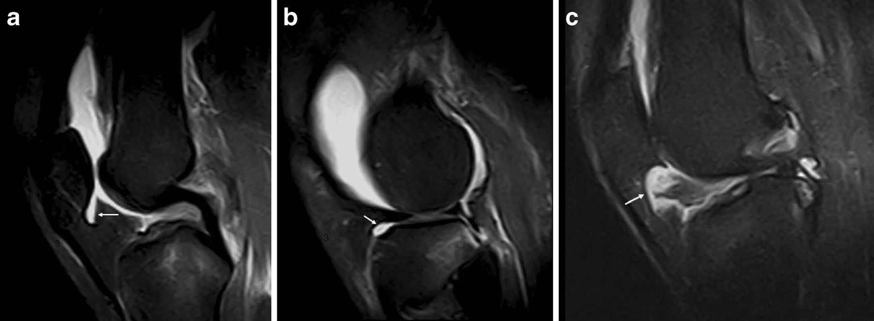
Sagittal proton-density fat-suppressed (PD FS) MRI demonstrates fluid in the suprapatellar pouch and fat-pad recesses. Arrows identify the suprahoffatic recess in a, the infrahoffatic recess in b, and a possible communication between the vertical and horizontal recesses in c.
Original Fig. 2. Draghi F, Ferrozzi G, Urciuoli L, Bortolotto C, Bianchi S. Hoffa’s fat pad abnormalities, knee pain and magnetic resonance imaging in daily practice. Insights Imaging. 2016;7(3):373–383. doi:10.1007/s13244-016-0483-8. © The Author(s) 2016. CC BY 4.0. Caption translated/adapted from original legend.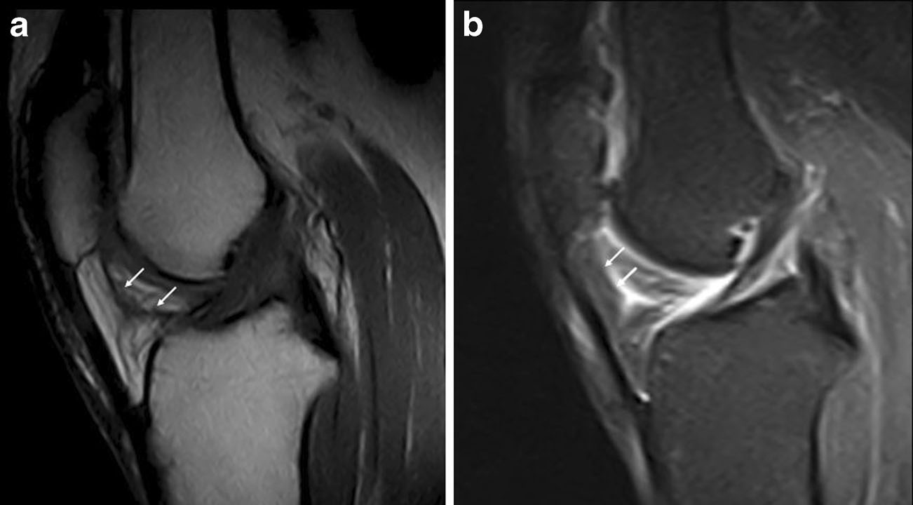
Sagittal T1-weighted (a) and PD FS (b) MR images demonstrate a thickened infrapatellar plica (arrows) with associated HFP edema. No age or additional clinical history is specified in the legend.
Original Fig. 3. Draghi F, Ferrozzi G, Urciuoli L, Bortolotto C, Bianchi S. Hoffa’s fat pad abnormalities, knee pain and magnetic resonance imaging in daily practice. Insights Imaging. 2016;7(3):373–383. doi:10.1007/s13244-016-0483-8. © The Author(s) 2016. CC BY 4.0. Caption translated/adapted from original legend.2|Superolateral edema and impingement: location matters, but so do symptoms
Repetitive microtrauma can inflame the fat pad. The review illustrates focal superolateral edema in a young athlete and an enlarged, edematous fat pad in a soccer player with Hoffa’s disease. With superolateral edema, review patellar height, the patellofemoral joint and adjacent patellar tendon. The review describes an association with patella alta, but edema alone does not establish a symptomatic impingement syndrome.
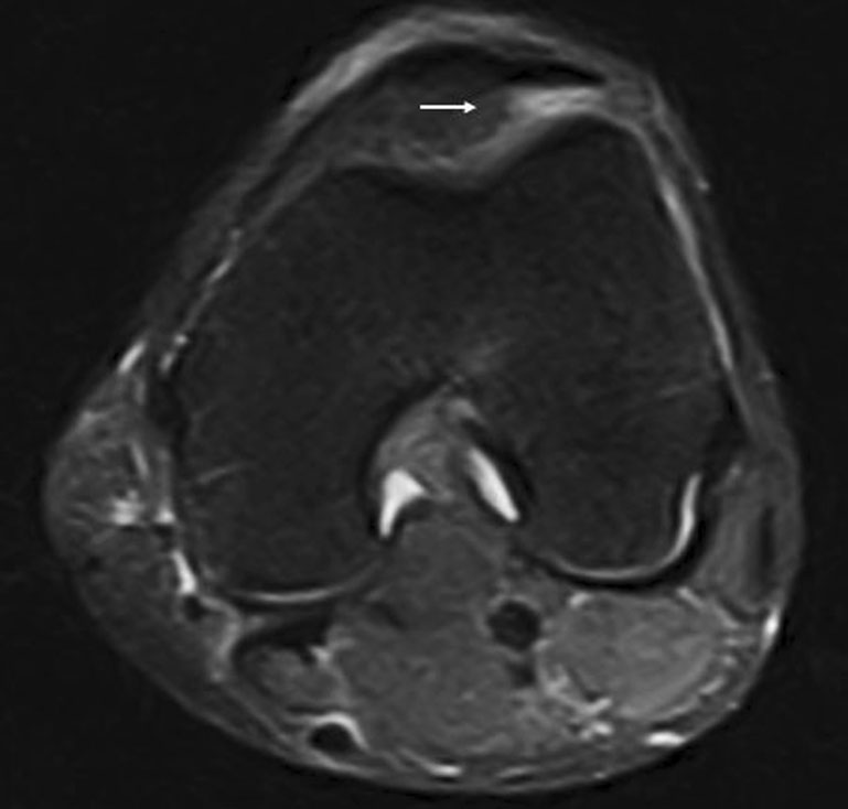
A young athlete with persistent superolateral knee pain. Axial PD FS MRI shows edema in the superolateral HFP, labeled superolateral HFP impingement in the source. The visible arrow lies at this region; the legend does not separately specify an arrow type.
Original Fig. 7. Draghi F, Ferrozzi G, Urciuoli L, Bortolotto C, Bianchi S. Hoffa’s fat pad abnormalities, knee pain and magnetic resonance imaging in daily practice. Insights Imaging. 2016;7(3):373–383. doi:10.1007/s13244-016-0483-8. © The Author(s) 2016. CC BY 4.0. Caption translated/adapted from original legend.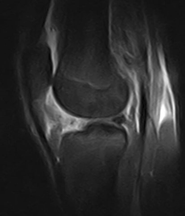
Hoffa’s disease in a soccer player exposed to repetitive hyperextension and rotational microtrauma. PD FS MRI shows an enlarged, edematous HFP. The original legend does not specify the plane or assign arrows or asterisks.
Original Fig. 8. Draghi F, Ferrozzi G, Urciuoli L, Bortolotto C, Bianchi S. Hoffa’s fat pad abnormalities, knee pain and magnetic resonance imaging in daily practice. Insights Imaging. 2016;7(3):373–383. doi:10.1007/s13244-016-0483-8. © The Author(s) 2016. CC BY 4.0. Caption translated/adapted from original legend.3|Acute trauma: read the fat pad in the context of the whole knee
Direct contusion, twisting injury and patellar dislocation may produce HFP edema or hemorrhage. Interpret increased signal on fluid-sensitive fat-suppressed sequences together with the patella, femoral condyles, tibial plateau, ligaments and joint fluid. Do not summarize a knee with substantial adjacent injury merely as fat-pad inflammation.
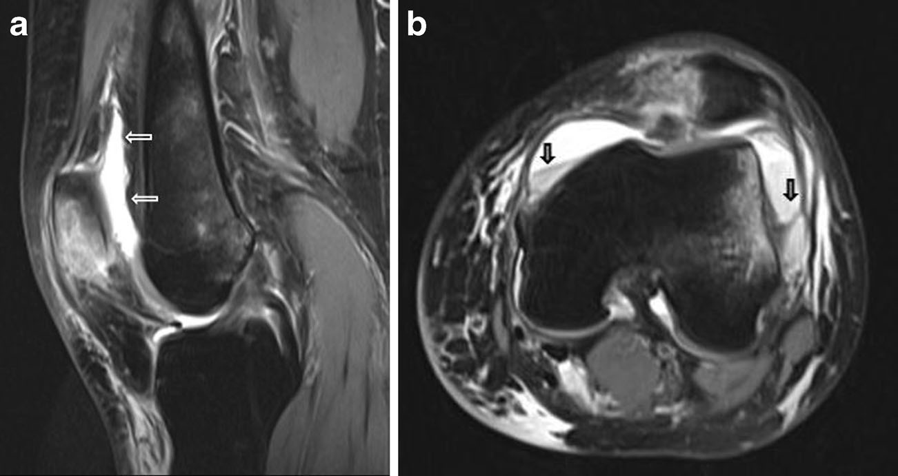
Acute direct contusion. PD FS images demonstrate HFP edema, patellar edema and joint effusion (a; thin arrows). Wide arrows on the axial image (b) mark fluid–fluid levels, described in the original legend as related to hemarthrosis.
Original Fig. 5. Draghi F, Ferrozzi G, Urciuoli L, Bortolotto C, Bianchi S. Hoffa’s fat pad abnormalities, knee pain and magnetic resonance imaging in daily practice. Insights Imaging. 2016;7(3):373–383. doi:10.1007/s13244-016-0483-8. © The Author(s) 2016. CC BY 4.0. Caption translated/adapted from original legend.4|Postoperative change: distinguish fibrosis from a Cyclops lesion
Establish whether the patient has undergone arthroscopy, anterior cruciate ligament (ACL) reconstruction or another operation. Fibrosis may appear as bands or ill-defined, confluent low-signal tissue. A Cyclops lesion is a nodular fibrous lesion after ACL reconstruction, typically anterior to the graft in the posterior HFP. In a patient with extension loss, its location and potential mechanical impingement deserve particular attention.
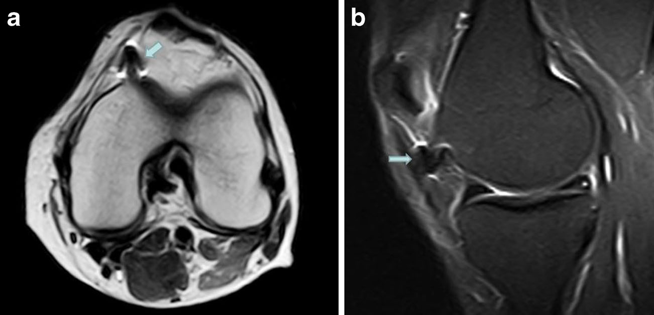
Post-surgical fibrosis. Axial T2-weighted (a) and sagittal PD FS (b) images show hypointense postoperative fibrotic tissue in the fat pad (arrow), with associated HFP edema.
Original Fig. 11. Draghi F, Ferrozzi G, Urciuoli L, Bortolotto C, Bianchi S. Hoffa’s fat pad abnormalities, knee pain and magnetic resonance imaging in daily practice. Insights Imaging. 2016;7(3):373–383. doi:10.1007/s13244-016-0483-8. © The Author(s) 2016. CC BY 4.0. Caption translated/adapted from original legend.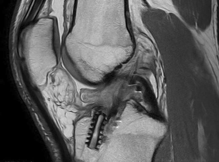
Cyclops lesion after ACL reconstruction. A sagittal proton-density image shows a hypointense mass within the edematous fat pad. The legend does not specify fat suppression or assign arrows or asterisks.
Original Fig. 10. Draghi F, Ferrozzi G, Urciuoli L, Bortolotto C, Bianchi S. Hoffa’s fat pad abnormalities, knee pain and magnetic resonance imaging in daily practice. Insights Imaging. 2016;7(3):373–383. doi:10.1007/s13244-016-0483-8. © The Author(s) 2016. CC BY 4.0. Caption translated/adapted from original legend.5|Trace the source: meniscus and patellar tendon
The HFP borders the menisci, ligaments and extensor mechanism, making secondary edema common. A low-signal structure within the pad may be a displaced meniscal flap rather than a primary mass. Patellar tendon thickening, increased signal or graft-harvest change can coexist with edema. In children and adolescents, also assess the tibial tubercle apophysis for Osgood–Schlatter disease.
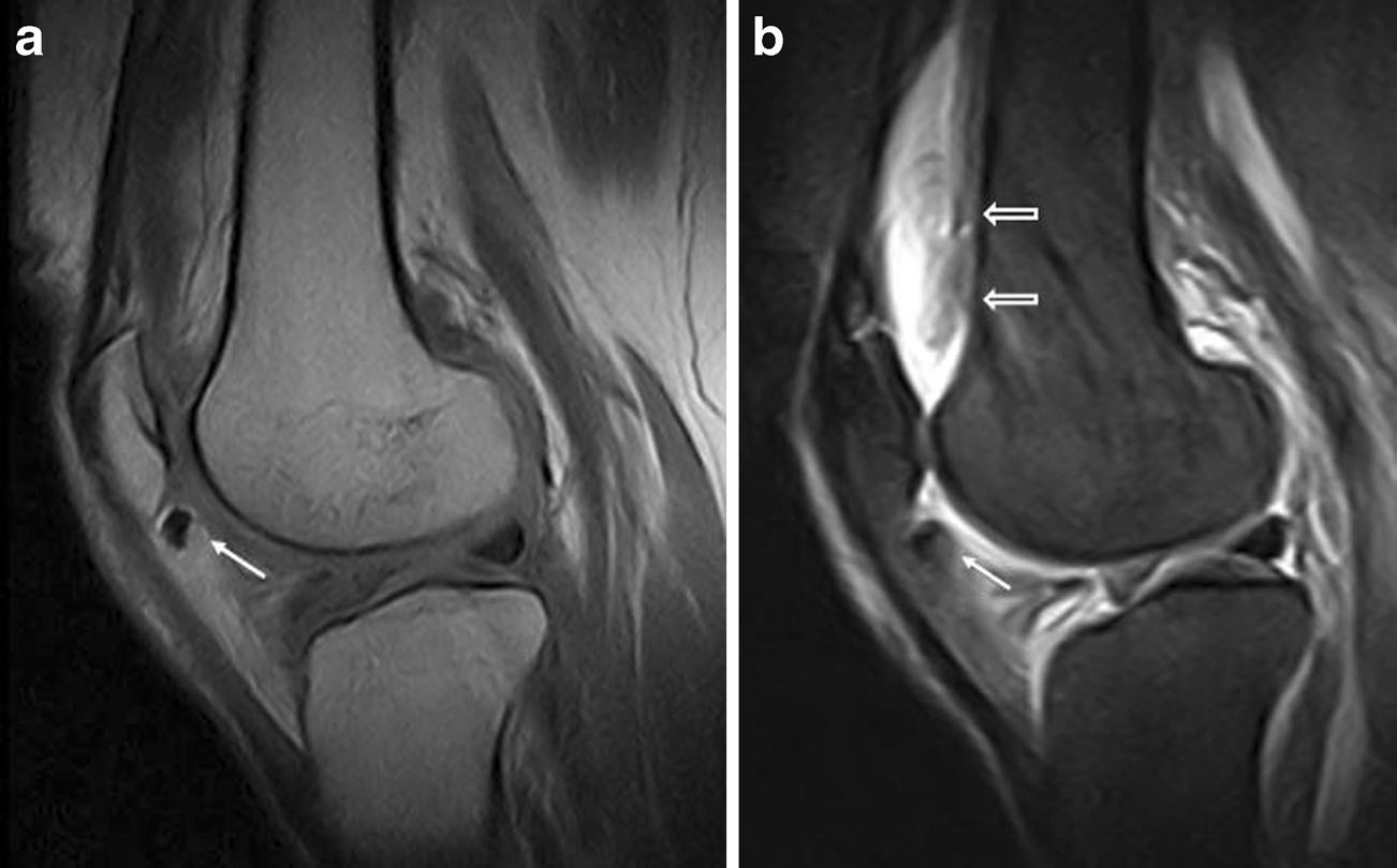
Lateral meniscal flap tear. Sagittal T1-weighted MRI (a) shows the displaced flap fragment within the fat pad (arrows). Sagittal PD FS MRI (b) demonstrates HFP edema, with joint effusion marked by wide arrows.
Original Fig. 12. Draghi F, Ferrozzi G, Urciuoli L, Bortolotto C, Bianchi S. Hoffa’s fat pad abnormalities, knee pain and magnetic resonance imaging in daily practice. Insights Imaging. 2016;7(3):373–383. doi:10.1007/s13244-016-0483-8. © The Author(s) 2016. CC BY 4.0. Caption translated/adapted from original legend.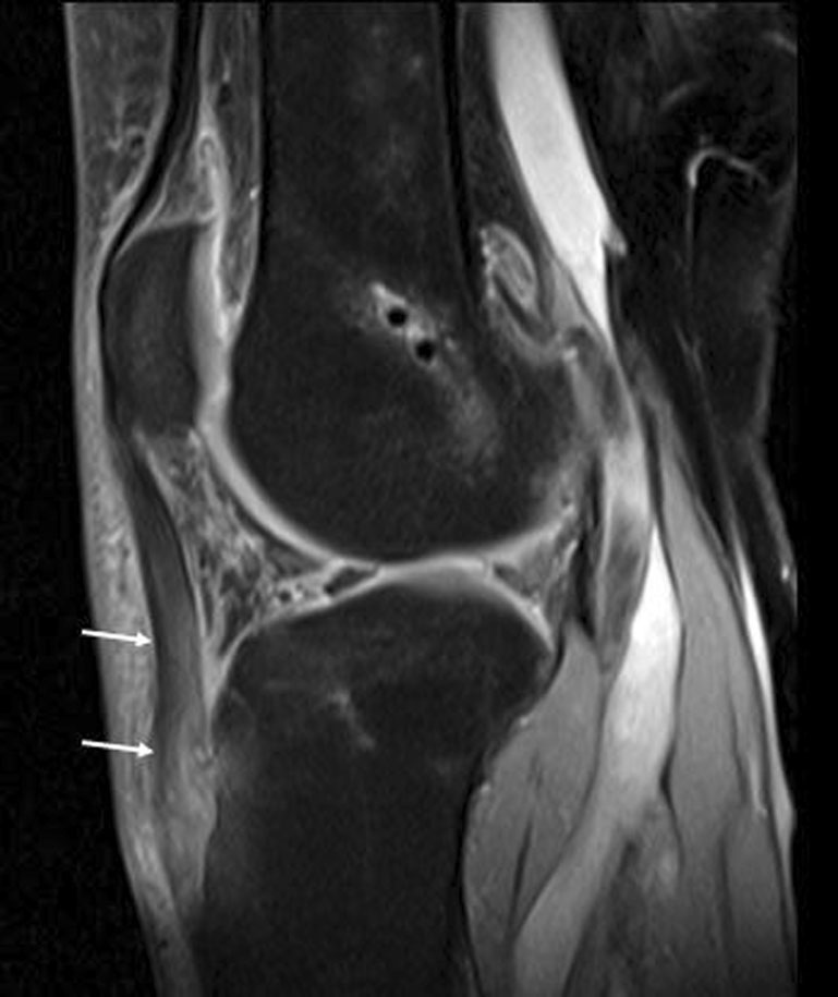
Patellar tendinopathy after autograft harvest. Sagittal PD FS MRI demonstrates increased patellar tendon signal and an increased anteroposterior diameter (arrows), with HFP edema.
Original Fig. 14. Draghi F, Ferrozzi G, Urciuoli L, Bortolotto C, Bianchi S. Hoffa’s fat pad abnormalities, knee pain and magnetic resonance imaging in daily practice. Insights Imaging. 2016;7(3):373–383. doi:10.1007/s13244-016-0483-8. © The Author(s) 2016. CC BY 4.0. Caption translated/adapted from original legend.6|Cystic lesions: meniscal cyst or ganglion?
When a fluid-signal lesion is found in the pad, trace its relationship to the meniscus and normal synovial recesses. Meniscal cysts commonly accompany a meniscal tear. Ganglia may be unilocular or multilocular, round or lobulated. If the adjacent meniscus appears normal, reconsider the origin instead of naming the lesion by location alone.
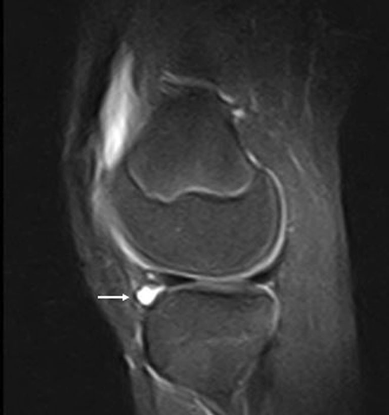
Meniscal cyst. A sagittal PD FS image (labeled a in the original legend) shows a cyst arising from the lateral meniscus and extending into the edematous fat pad (arrow).
Original Fig. 16. Draghi F, Ferrozzi G, Urciuoli L, Bortolotto C, Bianchi S. Hoffa’s fat pad abnormalities, knee pain and magnetic resonance imaging in daily practice. Insights Imaging. 2016;7(3):373–383. doi:10.1007/s13244-016-0483-8. © The Author(s) 2016. CC BY 4.0. Caption translated/adapted from original legend.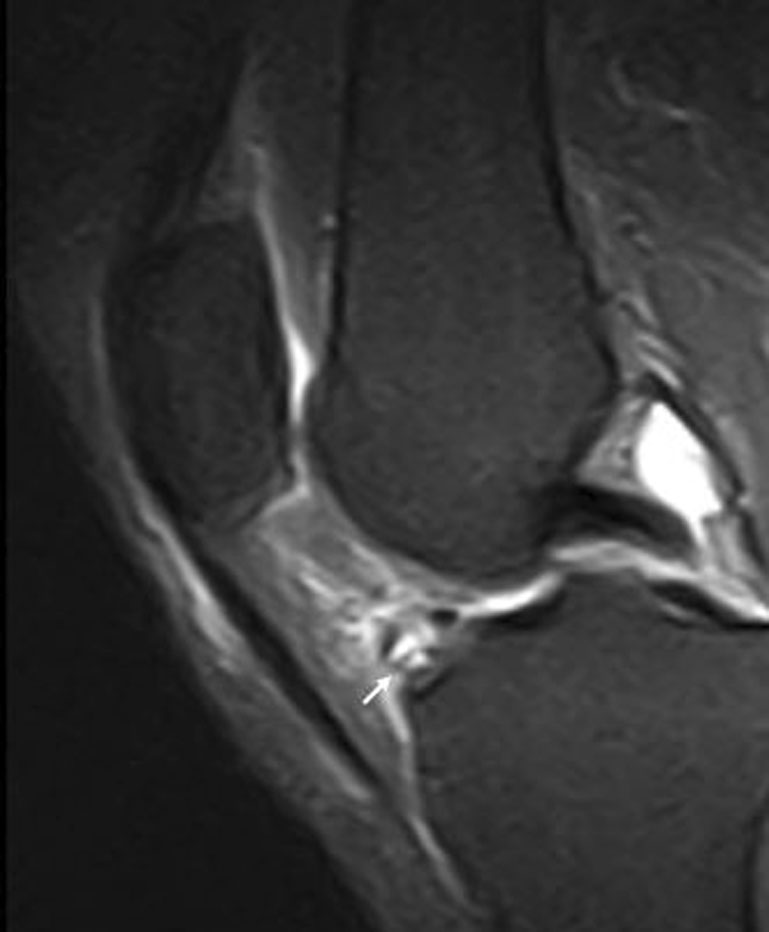
A patient with anterior knee pain and HFP edema. Sagittal PD FS MRI demonstrates a multilocular ganglion cyst within the fat pad (arrows). Age and surgical history are not specified in the legend.
Original Fig. 19. Draghi F, Ferrozzi G, Urciuoli L, Bortolotto C, Bianchi S. Hoffa’s fat pad abnormalities, knee pain and magnetic resonance imaging in daily practice. Insights Imaging. 2016;7(3):373–383. doi:10.1007/s13244-016-0483-8. © The Author(s) 2016. CC BY 4.0. Caption translated/adapted from original legend.7|Synovial disease: low-signal proliferation and loose bodies
When the abnormality extends beyond the fat pad, follow the synovium and joint recesses. The review uses pigmented villonodular synovitis (PVNS) to demonstrate hemosiderin-associated low-signal proliferation, and synovial chondromatosis to illustrate intra-articular loose bodies. Associated HFP edema should not distract from these more specific findings.
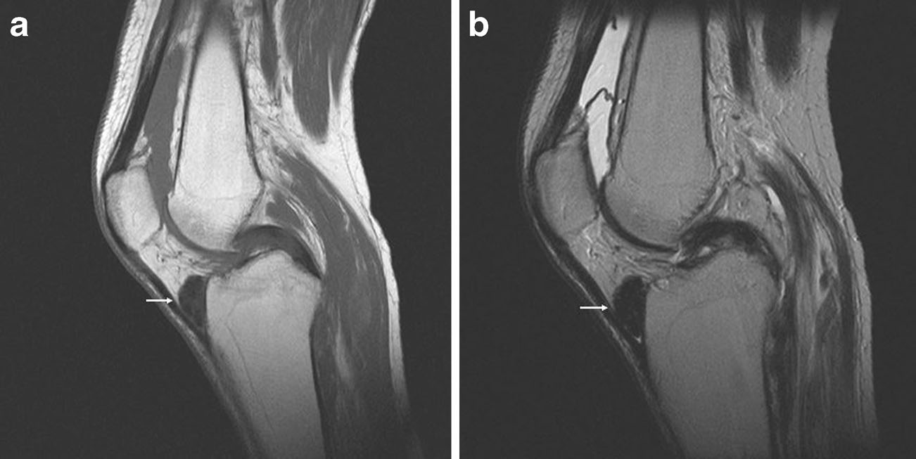
PVNS in the source terminology. Sagittal T1-weighted (a) and T2-weighted (b) images demonstrate synovial proliferations in the infrapatellar bursa (arrows). The legend attributes their hypointense signal to the paramagnetic effect of hemosiderin.
Original Fig. 17. Draghi F, Ferrozzi G, Urciuoli L, Bortolotto C, Bianchi S. Hoffa’s fat pad abnormalities, knee pain and magnetic resonance imaging in daily practice. Insights Imaging. 2016;7(3):373–383. doi:10.1007/s13244-016-0483-8. © The Author(s) 2016. CC BY 4.0. Caption translated/adapted from original legend.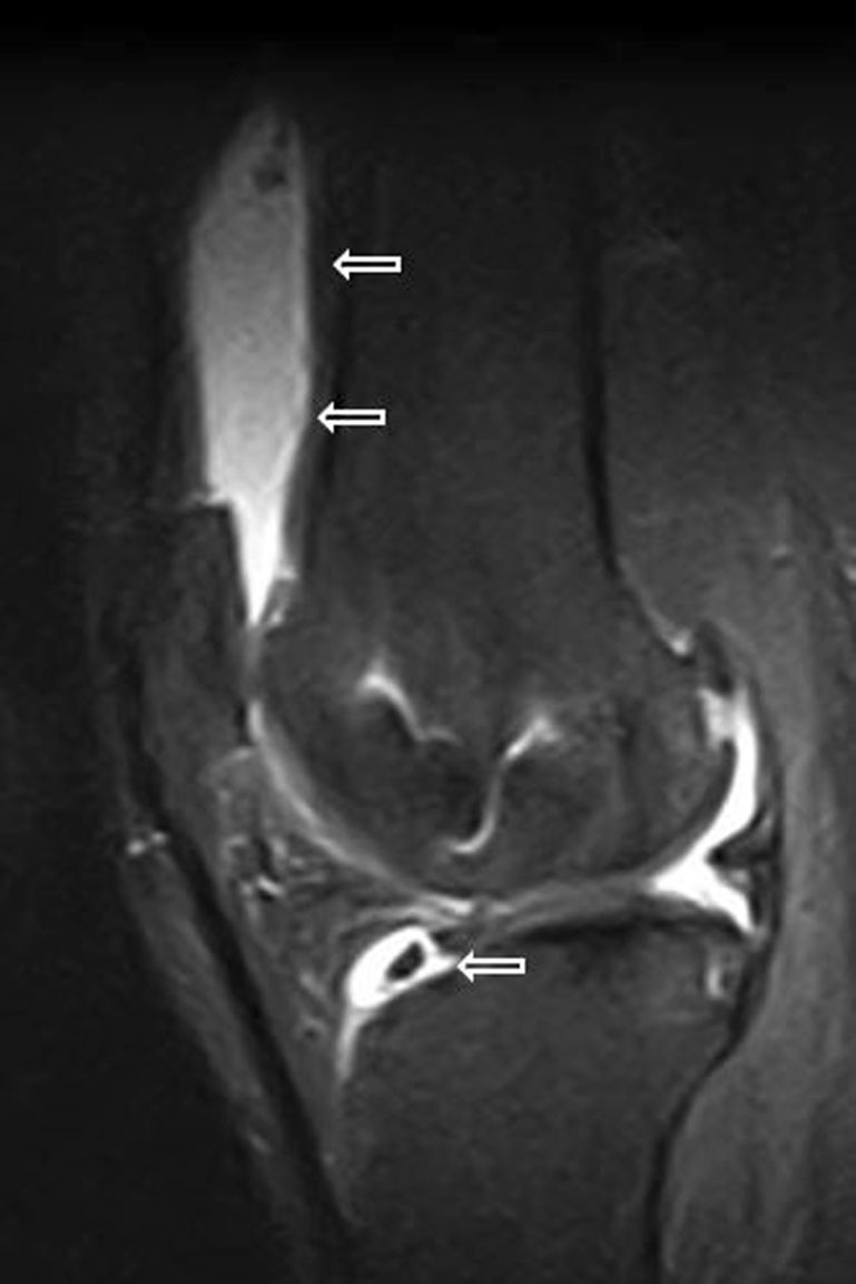
Synovial chondromatosis. Sagittal PD FS MRI shows joint effusion with loose bodies in the suprapatellar pouch and infrahoffatic recess (wide arrows), together with HFP edema.
Original Fig. 18. Draghi F, Ferrozzi G, Urciuoli L, Bortolotto C, Bianchi S. Hoffa’s fat pad abnormalities, knee pain and magnetic resonance imaging in daily practice. Insights Imaging. 2016;7(3):373–383. doi:10.1007/s13244-016-0483-8. © The Author(s) 2016. CC BY 4.0. Caption translated/adapted from original legend.Practical MRI Checklist
Take-home Messages
- HFP edema is an imaging finding, not proof of the pain generator.
- Recognize normal fat, recesses and plicae before diagnosing disease.
- Superolateral edema may suggest impingement-related change but requires clinical correlation.
- Describe postoperative fibrosis and Cyclops lesions separately, especially with extension loss.
- Trace the meniscus, tendon, ligaments and synovium to identify secondary involvement.
- Report location, morphology, primary pathology and clinical relevance rather than inferring treatment from edema alone.
Primary source and image license
Draghi F, Ferrozzi G, Urciuoli L, Bortolotto C, Bianchi S. Hoffa’s fat pad abnormalities, knee pain and magnetic resonance imaging in daily practice. Insights Imaging. 2016;7(3):373–383. doi:10.1007/s13244-016-0483-8.
© The Author(s) 2016. Creative Commons Attribution 4.0 International License (CC BY 4.0).
Original Figures 1, 2, 3, 7, 8, 5, 11, 10, 12, 14, 16, 19, 17 and 18 are reproduced from the embedded images in the supplied PDF. All panels and markers are retained without cropping, redrawing or recompression; the display of panel 1b is vertically scaled to align with a and c. Captions are translated/adapted from the original legends; teaching points and the reading pathway are an educational synthesis. No separate third-party credit line was found in the selected legends. Original Fig. 20 has a courtesy credit and is not reproduced here.
This page is for medical education and academic communication. It does not replace individual diagnosis or treatment advice.
Continue reading this category →