
Research & Academic Exchange
Putting Taiwanese Interventional Radiology on the Global Stage | TSIR × CIRSE Takeover: TAME for Refractory Ankle Osteoarthritis Pain
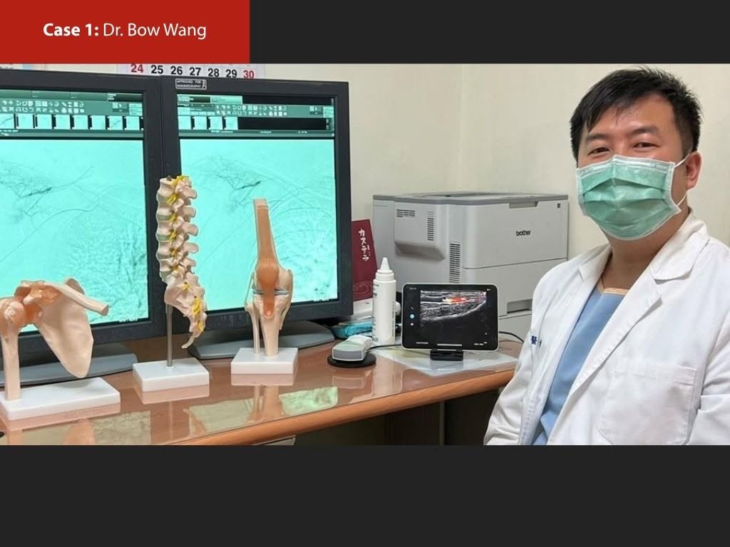
I was delighted to participate in the Taiwan Society of Interventional Radiology (TSIR) Takeover on CIRSE’s social platform and share a case of transarterial microembolization (TAME) for refractory pain associated with ankle osteoarthritis.
Using imaging to identify abnormal vascularity potentially involved in pain
The patient was a 61-year-old man with left ankle osteoarthritis. Severe pain persisted despite medication, rehabilitation, injections, and arthroscopic debridement. His baseline numerical rating scale (NRS) pain score was 9.
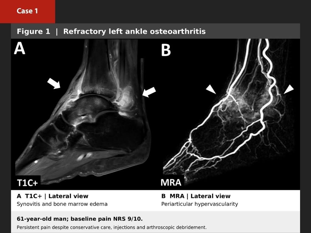
Preprocedural contrast-enhanced MRI demonstrated synovitis and subchondral bone marrow edema, while MR angiography showed abnormal periarticular hypervascularity. These findings must be interpreted together with the history, pain location, and physical examination before planning treatment.
Selective treatment of abnormal vascularity via the anterior tibial artery
Angiography of the anterior tibial artery demonstrated abnormal periarticular vascularity corresponding to the painful region. After selective catheter positioning, embolization was performed with imipenem/cilastatin. Postembolization angiography showed marked elimination of the abnormal blush while preserving normal arterial supply.
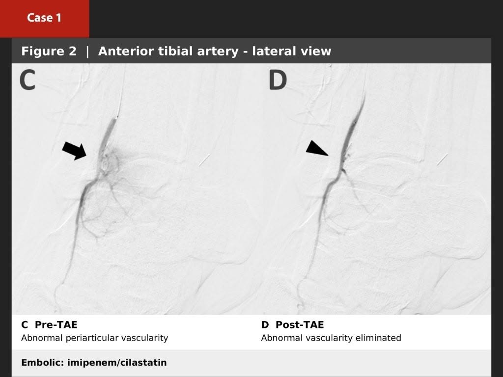
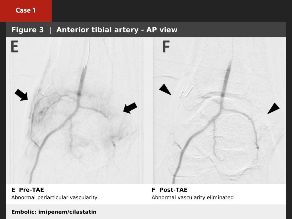
The patient’s pain decreased from an NRS score of 9 at baseline to 3 at six months. This is an individual clinical case and should not be interpreted as evidence that every patient with ankle osteoarthritis will experience the same result.
TAME is more than “blocking a vessel”
The central message is not simply the technical act of embolization. It is the integration of clinical history, MRI, MR angiography, catheter angiography, and the precise pain location to identify abnormal vascularity that may contribute to chronic pain and to deliver individualized treatment.
TAME is not intended to eliminate the normal blood supply of the ankle. The procedure requires selective identification of abnormal vascular signals, careful reduction of nontarget embolization risk, and continued follow-up of pain, function, and imaging findings.
TSIR × CIRSE: bringing Taiwan’s experience to the international community
TSIR has long supported the clinical development, professional education, and international exchange of interventional radiology in Taiwan. CIRSE—the Cardiovascular and Interventional Radiological Society of Europe—is a major international professional society in cardiovascular and interventional radiology. TSIR formally became a CIRSE group member in 2026, creating additional opportunities for Taiwanese interventional radiologists to participate in international education, research, and professional exchange.
My special thanks go to TSIR President Dr. Pi-Yi Chang for planning and advancing this exchange, enabling Taiwan’s experience in pain intervention and musculoskeletal embolization to reach more international colleagues.
I was also pleased that this Takeover featured phantom-limb pain cases from Dr. Tien-Yu Wu and Dr. Cheng-Han Huang of Shin Kong Wu Ho-Su Memorial Hospital. Together, these cases illustrate how interventional radiology is expanding beyond its traditional roles in oncology and vascular disease into chronic pain and musculoskeletal conditions.
Further reading
CIRSE Takeover: Bow Wang’s TAME case for ankle osteoarthritis pain
TSIR × CIRSE Takeover event post
CIRSE: New group member — Taiwan Society of Interventional Radiology
This page is for medical education and academic communication. It does not replace individual diagnosis or treatment advice.
Continue reading this category →