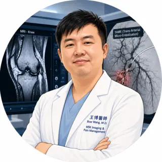
Research & Academic Exchange
Transarterial Microembolization (TAME) for Chronic Musculoskeletal Pain: Notes from Kaohsiung
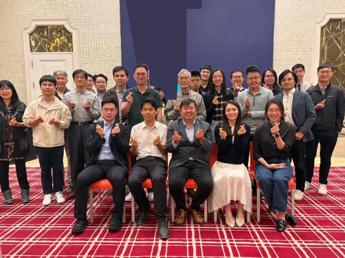
From our hospital’s experience to a case of persistent wrist pain and a discussion of what to reassess when treatment falls short, this meeting brought radiologists and cardiologists together to compare how they approach TAME.
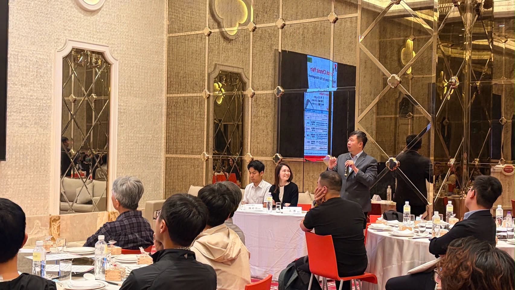
On November 14, I joined Unlock Chronic Pain: Next-Gen Solutions for Pain Management in Kaohsiung, organized by ASAHI INTECC. The meeting focused on transarterial microembolization (TAME) for chronic musculoskeletal pain. I gave the opening remarks and served as a moderator and panelist alongside Dr. Sz-Tsan Wang (王思讚) and Dr. Jih-Tze Tan (陳祈池), both from Dalin Tzu Chi Hospital.
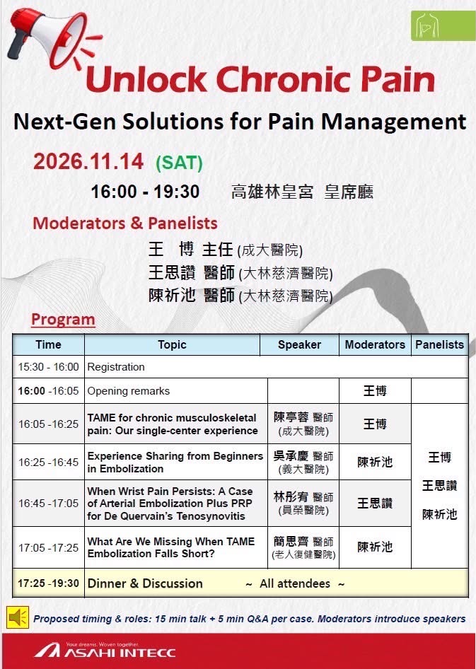
Four talks: From clinical experience to what to revisit when TAME falls short
- Dr. Ting-Rong Chen (陳亭蓉, National Cheng Kung University Hospital) presented our single-center experience with TAME for chronic musculoskeletal pain. I moderated the session.
- Dr. Cheng-Ching Wu (吳承慶, E-Da Hospital) shared lessons from getting started with embolization. Dr. Jih-Tze Tan moderated.
- Dr. Tung-Yu Lin (林彤宥, Yuan-Rung Hospital) presented a case of persistent wrist pain due to de Quervain’s tenosynovitis and discussed arterial embolization. Dr. Sz-Tsan Wang moderated.
- Dr. Szu-Chi Chien (簡思齊, Taichung Municipal Geriatric Rehabilitation General Hospital) discussed what to reassess when TAME falls short. Dr. Jih-Tze Tan moderated.
The final talk brought me back to what I consider the most important question: If pain hasn’t improved after TAME, the first step is to revisit whether the initial diagnosis was correct. Does the source of pain we identified still explain the patient’s symptoms? Answering that question gives us a clearer starting point for reviewing the angiogram and the embolization procedure.
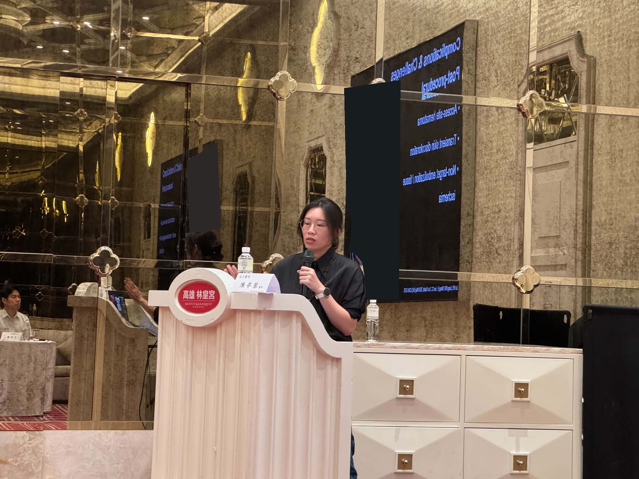
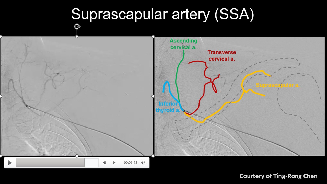
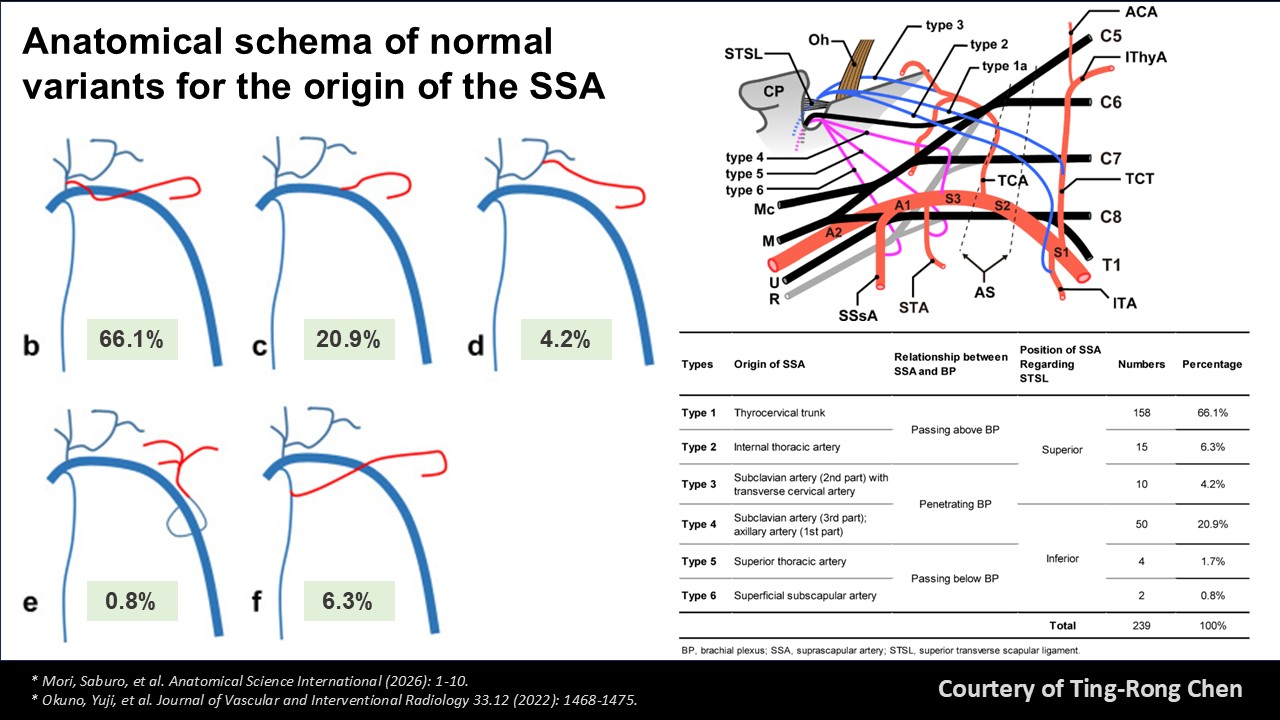
Reading angiograms for TAME: The suprascapular artery
In her talk on our hospital’s experience with TAME, Dr. Ting-Rong Chen used the two slides below to illustrate how to identify the suprascapular artery (SSA). The first uses color to trace the branches on an angiogram. The second shows how the artery can take different routes from different points of origin. In the anatomical study by Mori and colleagues, the most common origin was the thyrocervical trunk, found in 158 of 239 arteries (66.1%). That percentage describes the study sample. When reading an individual angiogram, we still need to trace where the artery begins and where it goes.
The meeting reinforced this point for me: when pain hasn’t improved, the priority is to revisit the initial diagnosis. I would ask the patient again where and when it hurts and which movement brings on the pain, then put those answers together with the physical examination and imaging to see whether the original explanation still fits.
My thanks to ASAHI INTECC for organizing the meeting and bringing radiologists and cardiologists together to share their experience with TAME. Thank you also to the speakers, moderators, and everyone who brought their clinical questions to the discussion.
Further reading: Mori et al. on the origin and course of the suprascapular artery; Okuno et al.’s feasibility study of embolization for refractory nighttime shoulder pain.
This page is for medical education and academic communication. It does not replace individual diagnosis or treatment advice.
Continue reading this category →